Anti-Calsequestrin antibody (ab3516)
")
Key features and details
- Rabbit polyclonal to Calsequestrin 2 + Calsequestrin 1
- Suitable for: ICC/IF, WB, IP, IHC-P
- Reacts with: Mouse, Rat, Sheep, Rabbit, Dog, Human, Pig, Rainbow trout
- Isotype: IgG
Overview
-
Product name
Anti-Calsequestrin 2 + Calsequestrin 1 antibody -
Description
Rabbit polyclonal to Calsequestrin 2 + Calsequestrin 1 -
Host species
Rabbit -
Specificity
This antibody recognizes both cardiac and skeletal muscle calsequestrin. -
Tested applications
Suitable for: ICC/IF, WB, IP, IHC-Pmore details -
Species reactivity
Reacts with: Mouse, Rat, Sheep, Rabbit, Dog, Human, Pig, Rainbow trout -
Immunogen
Full length native protein (purified) corresponding to Dog Calsequestrin 2. The immunogen is purified canine cardiac calsequestrin.
-
General notes
The Life Science industry has been in the grips of a reproducibility crisis for a number of years. Abcam is leading the way in addressing this with our range of recombinant monoclonal antibodies and knockout edited cell lines for gold-standard validation. Please check that this product meets your needs before purchasing.
If you have any questions, special requirements or concerns, please send us an inquiry and/or contact our Support team ahead of purchase. Recommended alternatives for this product can be found below, along with publications, customer reviews and Q&As
Properties
-
Form
Liquid -
Storage instructions
Shipped at 4°C. Store at +4°C short term (1-2 weeks). Upon delivery aliquot. Store at -20°C or -80°C. Avoid freeze / thaw cycle. -
Storage buffer
Preservative: 0.05% Sodium azide -
 Concentration information loading...
 -
Purity
Whole antiserum -
Clonality
Polyclonal -
Isotype
IgG -
Research areas
Images
-
Western blot - Anti-Calsequestrin antibody (ab3516)All lanes : Anti-Calsequestrin antibody (ab3516) at 1/5000 dilution
Lane 1 : RD (Human muscle rhabdomyosarcoma cell line) whole cell lysate
Lane 2 : L6 (Rat skeletal muscle cell line) whole cell lysate
Lane 3 : Mouse heart tissue lysate
Lysates/proteins at 25 µg per lane.
Secondary
All lanes : HRP-conjugated secondary antibody
Observed band size: 55 kDa why is the actual band size different from the predicted?
-
 Immunohistochemistry (Formalin/PFA-fixed paraffin-embedded sections) - Anti-Calsequestrin antibody (ab3516)
ab3516 labelling Calsequestrin in the cytoplasm of human skeletal muscle tissue (right) compared with a negative control (left) by Immunohistochemistry (formalin/PFA-fixed paraffin-embedded sections). To expose target proteins, antigen retrieval method was performed using 10mM sodium citrate (pH 6.0) microwaved for 8-15 min. Tissues were blocked in 3% H2O2-methanol for 15 min at room temperature, washed with ddH2O and PBS. Tissue sections were incubated with primary antibody (1:200 in 3% BSA-PBS) overnight at 4°C. A HRP-conjugated anti-rabbit was used as the secondary antibody, followed by colorimetric detection using a DAB kit. Tissues were counterstained with hematoxylin and dehydrated with ethanol and xylene to prep for mounting.
-
 Immunocytochemistry/ Immunofluorescence - Anti-Calsequestrin antibody (ab3516)
Immunofluorescent analysis of Calsequestrin (green) showing staining in the cytoplasm and nucleus of C2C12 (Mouse myoblast cell line) cells (right) compared to a negative control without primary antibody (left). Formalin-fixed cells were permeabilized with 0.1% Triton X-100 in TBS for 5-10 minutes and blocked with 3% BSA-PBS for 30 minutes at room temperature. Cells were probed with ab3516 in 3% BSA-PBS at a dilution of 1/100 and incubated overnight at 4 °C in a humidified chamber. Cells were washed with PBST and incubated with a DyLight-conjugated secondary antibody in PBS at room temperature in the dark. F-actin (red) was stained with a fluorescent red phalloidin and nuclei (blue) were stained with Hoechst or DAPI. Images were taken at a magnification of 60x.
-
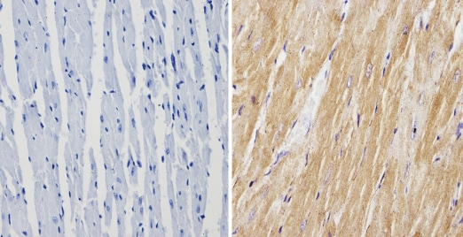 Immunohistochemistry (Formalin/PFA-fixed paraffin-embedded sections) - Anti-Calsequestrin antibody (ab3516)
ab3516 labelling Calsequestrin in the cytoplasm of mouse heart tissue (right) compared with a negative control (left) by Immunohistochemistry (formalin/PFA-fixed paraffin-embedded sections). To expose target proteins, antigen retrieval method was performed using 10mM sodium citrate (pH 6.0) microwaved for 8-15 min. Following antigen retrieval, tissues were blocked in 3% H2O2-methanol for 15 min at room temperature, washed with ddH2O and PBS. Tissue sections were incubated with primary antibody (1:200 in 3% BSA-PBS) overnight at 4°C. A HRP-conjugated anti-rabbit was used as the secondary antibody, followed by colorimetric detection using a DAB kit. Tissues were counterstained with hematoxylin and dehydrated with ethanol and xylene to prep for mounting.
-
 Immunohistochemistry (Formalin/PFA-fixed paraffin-embedded sections) - Anti-Calsequestrin antibody (ab3516)
ab3516 labelling Calsequestrin in the cytoplasm of human heart tissue (right) compared with a negative control (left) by Immunohistochemistry (formalin/PFA-fixed paraffin-embedded sections). To expose target proteins, antigen retrieval method was performed using 10mM sodium citrate (pH 6.0) microwaved for 8-15 min. Following antigen retrieval, tissues were blocked in 3% H2O2-methanol for 15 min at room temperature, washed with ddH2O and PBS. Tissue sections were incubated with primary antibody (1:200 in 3% BSA-PBS) overnight at 4°C. A HRP-conjugated anti-rabbit was used as the secondary antibody, followed by colorimetric detection using a DAB kit. Tissues were counterstained with hematoxylin and dehydrated with ethanol and xylene to prep for mounting.
-
 Immunohistochemistry (Formalin/PFA-fixed paraffin-embedded sections) - Anti-Calsequestrin antibody (ab3516)
ab3516 (1µg/ml) staining Calsequestrin in human skeletal muscle using an automated system (DAKO Autostainer Plus). Using this protocol there is strong staining of discrete organelles within the cytoplasm.
Sections were rehydrated and antigen retrieved with the Dako 3 in 1 AR buffer EDTA pH 9.0 in a DAKO PT link. Slides were peroxidase blocked in 3% H2O2 in methanol for 10 mins. They were then blocked with Dako Protein block for 10 minutes (containing casein 0.25% in PBS) then incubated with primary antibody for 20 min and detected with Dako envision flex amplification kit for 30 minutes. Colorimetric detection was completed with Diaminobenzidine for 5 minutes. Slides were counterstained with Haematoxylin and coverslipped under DePeX. Please note that, for manual staining, optimization of primary antibody concentration and incubation time is recommended. Signal amplification may be required. -
 Immunocytochemistry/ Immunofluorescence - Anti-Calsequestrin antibody (ab3516) This image is courtesy of an anonymous Abreview
ab3516 staining Calsequestrin in mouse myocyte cell by ICC/IF (Immunocytochemistry/immunofluorescence). Cells were fixed with formaldehyde and blocked with 10% serum for 1 hour at 25°C. Samples were incubated with primary antibody (1/50 in serum) for 18 hours at 4°C. An Alexa Fluor®488-conjugated Goat anti-rabbit IgG polyclonal (1/1000) was used as the secondary antibody.