Anti-beta COP antibody (ab24359)
")
Key features and details
- Rabbit polyclonal to beta COP
- Suitable for: WB, ICC/IF
- Reacts with: Mouse, Rat
- Isotype: IgG
Overview
-
Product name
Anti-beta COP antibody
See all beta COP primary antibodies -
Description
Rabbit polyclonal to beta COP -
Host species
Rabbit -
Tested applications
Suitable for: WB, ICC/IFmore details -
Species reactivity
Reacts with: Mouse, Rat
Predicted to work with: Chicken, Human, Zebrafish
-
Immunogen
Synthetic peptide. This information is proprietary to Abcam and/or its suppliers.
-
Positive control
- Rat and mouse brain lysate.
-
General notes
The Life Science industry has been in the grips of a reproducibility crisis for a number of years. Abcam is leading the way in addressing this with our range of recombinant monoclonal antibodies and knockout edited cell lines for gold-standard validation. Please check that this product meets your needs before purchasing.
If you have any questions, special requirements or concerns, please send us an inquiry and/or contact our Support team ahead of purchase. Recommended alternatives for this product can be found below, along with publications, customer reviews and Q&As
Properties
-
Form
Liquid -
Storage instructions
Shipped at 4°C. Store at +4°C short term (1-2 weeks). Upon delivery aliquot. Store at -20°C or -80°C. Avoid freeze / thaw cycle. -
Storage buffer
pH: 7.40
Preservative: 0.02% Sodium azide
Constituent: PBS
Batches of this product that have a concentration Concentration information loading...
Purity
Immunogen affinity purifiedClonality
PolyclonalIsotype
IgGResearch areas
Associated products
-
Compatible Secondaries
-
Isotype control
-
Positive Controls
Applications
The Abpromise guarantee
Our Abpromise guarantee covers the use of ab24359 in the following tested applications.
The application notes include recommended starting dilutions; optimal dilutions/concentrations should be determined by the end user.
Application Abreviews Notes WB Use a concentration of 1 µg/ml. Detects a band of approximately 100 kDa (predicted molecular weight: 110 kDa).ICC/IF Use a concentration of 1 µg/ml.Notes WB
Use a concentration of 1 µg/ml. Detects a band of approximately 100 kDa (predicted molecular weight: 110 kDa).ICC/IF
Use a concentration of 1 µg/ml.Target
-
Function
The coatomer is a cytosolic protein complex that binds to dilysine motifs and reversibly associates with Golgi non-clathrin-coated vesicles, which further mediate biosynthetic protein transport from the ER, via the Golgi up to the trans Golgi network. Coatomer complex is required for budding from Golgi membranes, and is essential for the retrograde Golgi-to-ER transport of dilysine-tagged proteins. In mammals, the coatomer can only be recruited by membranes associated to ADP-ribosylation factors (ARFs), which are small GTP-binding proteins; the complex also influences the Golgi structural integrity, as well as the processing, activity, and endocytic recycling of LDL receptors. Plays a functional role in facilitating the transport of kappa-type opioid receptor mRNAs into axons and enhances translation of these proteins. Required for limiting lipid storage in lipid droplets. Involved in lipid homeostasis by regulating the presence of perilipin family members PLIN2 and PLIN3 at the lipid droplet surface and promoting the association of adipocyte surface triglyceride lipase (PNPLA2) with the lipid droplet to mediate lipolysis (By similarity). Involved in the Golgi disassembly and reassembly processes during cell cycle. Involved in autophagy by playing a role in early endosome function. Plays a role in organellar compartmentalization of secretory compartments including endoplasmic reticulum (ER)-Golgi intermediate compartment (ERGIC), Golgi, trans-Golgi network (TGN) and recycling endosomes, and in biosynthetic transport of CAV1. Promotes degradation of Nef cellular targets CD4 and MHC class I antigens by facilitating their trafficking to degradative compartments. -
Sequence similarities
Contains 6 HEAT repeats. -
Post-translational
modificationsProteolytically cleaved between Ser-528 and Ser-529 by CAPN8. -
Cellular localization
Cytoplasm. Golgi apparatus membrane. Cytoplasmic vesicle > COPI-coated vesicle membrane. Cell membrane. Endoplasmic reticulum-Golgi intermediate compartment. The coatomer is cytoplasmic or polymerized on the cytoplasmic side of the Golgi, as well as on the vesicles/buds originating from it. Proteolytic cleavage by CAPN8 triggers translocation from Golgi to cytoplasm (By similarity). Found in perinuclear vesicular-tubular clusters (VTCs) and in the Golgi region where associated with vesicles, buds and rims of the Golgi stack (By similarity). Occasionally present at the trans-side of Golgi, but mainly present at the cis-Golgi side in transitional areas (TA), on so-called peripheral elements (PE) consisting of tubules and vesicles located between the cup-shaped transitional elements (TE) of the rough endoplasmic reticulum (RER) and the cis-most Golgi cisternae (By similarity). Present in cytoplasm, not associated with visible coats or membranes, with a minor fraction present on small clusters of tubules and vesicles (By similarity). Some association with high-density and low-density microsomes and mitochondria/nuclei fraction (By similarity). Very little found in plasma membrane fraction. - Information by UniProt
-
Database links
- Entrez Gene: 423063 Chicken
- Entrez Gene: 1315 Human
- Entrez Gene: 70349 Mouse
- Entrez Gene: 114023 Rat
- Entrez Gene: 338138 Zebrafish
- Omim: 600959 Human
- SwissProt: Q5ZIA5 Chicken
- SwissProt: P53618 Human
see all -
Alternative names
- Beta-coat protein antibody
- Beta-COP antibody
- betacop antibody
see all
Images
-
Western blot - Anti-beta COP antibody (ab24359)All lanes : Anti-beta COP antibody (ab24359) at 1 µg/ml
Lane 1 : Mouse brain lysate
Lane 2 : Rat brain lysate
Lysates/proteins at 20 µg per lane.
Secondary
All lanes : Goat polyclonal to Rabbit IgG (Alexa Fluor® 680) at 1/10000 dilution
Performed under reducing conditions.
Predicted band size: 110 kDa
Observed band size: 100 kDa why is the actual band size different from the predicted?ab24359 detected a band of ~ 100 kDa in mouse and rat brain lysate. It also detected a band of ~ 100 kDa in NIH 3T3 cells and rat liver cells (data not shown). ab24359 is predicted to react with human beta cop due to 100% identity with the immunogen, however in our hands the antibody failed to detect a band in human brain lysate.
-
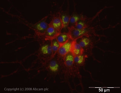 Immunocytochemistry/ Immunofluorescence - Anti-beta COP antibody (ab24359)
ICC/IF image of ab24359 stained rat PC12 cells. The cells were PFA fixed (10 min), permabilised in PBS-T (20 min) and incubated with the antibody (ab24359, 1µg/ml) for 1h at room temperature. 1%BSA / 10% normal goat serum / 0.3M glycine was used to block non-specific protein-protein interactions. The secondary antibody (green) was Alexa Fluor® 488 goat anti-rabbit IgG (H+L) used at a 1/1000 dilution for 1h. Alexa Fluor® 594 WGA was used to label plasma membranes (red). DAPI was used to stain the cell nuclei (blue).
Protocols
Datasheets and documents
-
SDS download
-
Datasheet download
References (2)
ab24359 has been referenced in 2 publications.
- Howley BV et al. A CREB3-regulated ER-Golgi trafficking signature promotes metastatic progression in breast cancer. Oncogene 37:1308-1325 (2018). PubMed: 29249802
- Fétaud-Lapierre V et al. Time-course proteomic analysis of taurocholate-induced necrotizing acute pancreatitis. J Proteomics 85:12-27 (2013). PubMed: 23624238
Images
-
Western blot - Anti-beta COP antibody (ab24359)All lanes : Anti-beta COP antibody (ab24359) at 1 µg/ml
Lane 1 : Mouse brain lysate
Lane 2 : Rat brain lysate
Lysates/proteins at 20 µg per lane.
Secondary
All lanes : Goat polyclonal to Rabbit IgG (Alexa Fluor® 680) at 1/10000 dilution
Performed under reducing conditions.
Predicted band size: 110 kDa
Observed band size: 100 kDa why is the actual band size different from the predicted?ab24359 detected a band of ~ 100 kDa in mouse and rat brain lysate. It also detected a band of ~ 100 kDa in NIH 3T3 cells and rat liver cells (data not shown). ab24359 is predicted to react with human beta cop due to 100% identity with the immunogen, however in our hands the antibody failed to detect a band in human brain lysate.
-
Immunocytochemistry/ Immunofluorescence - Anti-beta COP antibody (ab24359)
ICC/IF image of ab24359 stained rat PC12 cells. The cells were PFA fixed (10 min), permabilised in PBS-T (20 min) and incubated with the antibody (ab24359, 1µg/ml) for 1h at room temperature. 1%BSA / 10% normal goat serum / 0.3M glycine was used to block non-specific protein-protein interactions. The secondary antibody (green) was Alexa Fluor® 488 goat anti-rabbit IgG (H+L) used at a 1/1000 dilution for 1h. Alexa Fluor® 594 WGA was used to label plasma membranes (red). DAPI was used to stain the cell nuclei (blue).


