Anti-alpha Tubulin antibody - Microtubule Marker (ab18251)
")
Key features and details
- Rabbit polyclonal to alpha Tubulin - Microtubule Marker
- Suitable for: ICC/IF, Flow Cyt, WB
- Reacts with: Mouse, Rat, Human
- Isotype: IgG
Overview
-
Product name
Anti-alpha Tubulin antibody - Microtubule Marker
See all alpha Tubulin primary antibodies -
Description
Rabbit polyclonal to alpha Tubulin - Microtubule Marker -
Host species
Rabbit -
Tested Applications & Species
See all applications and species dataApplication Species Flow Cyt MouseRatHumanICC/IF MouseRatHumanWB MouseRatHuman
-
Immunogen
Synthetic peptide conjugated to KLH derived from within residues 400 to the C-terminus of Human alpha Tubulin.
Read Abcam's proprietary immunogen policy -
Positive control
- WB: HeLa, HEK-293, HepG2, Caco-2, NIH/3T3 and PC-12 whole cell lysates. ICC/IF: HeLa, Caco-2, NIH/3T3 and SV40LT-SMC cells.
Images
-
Western blot - Anti-alpha Tubulin antibody - Microtubule Marker (ab18251)All lanes : Anti-alpha Tubulin antibody - Microtubule Marker (ab18251) at 0.5 µg/ml
Lane 1 : HeLa (Human epithelial carcinoma cell line) whole cell lysate
Lane 2 : HEK-293 (Human embryonic kidney cell line) whole cell lysate
Lane 3 : HepG2 (Human hepatocellular liver carcinoma cell line) whole cell lysate
Lane 4 : Caco-2 (Human colonic carcinoma cell line) whole cell lysate
Lane 5 : NIH/3T3 (Mouse embryonic fibroblast cell line) whole cell lysate
Lane 6 : PC-12 (Rat adrenal pheochromocytoma cell line) whole cell lysate
Lysates/proteins at 20 µg per lane.
Secondary
All lanes : Goat Anti-Rabbit IgG H&L (Alexa Fluor® 790) (ab175781) at 1/10000 dilution
Predicted band size: 50 kDa
Observed band size: 52 kDa why is the actual band size different from the predicted?This blot was produced using a 4-12% Bis-tris gel under the MOPS buffer system. The gel was run at 200V for 50 minutes before being transferred onto a Nitrocellulose membrane at 30V for 70 minutes. The membrane was then blocked for an hour using Licor blocking buffer before being incubated with ab18251 overnight at 4°C. Antibody binding was detected using Anti-Rabbit Alexa Fluor® 790 (ab175781) at a 1:10,000 dilution for 1hr at room temperature and then imaged using the Licor Odyssey CLx.
-
 Immunocytochemistry/ Immunofluorescence - Anti-alpha Tubulin antibody - Microtubule Marker (ab18251)
ab18251 staining alpha-Tubulin in SV40LT-SMC cells.
The cells were fixed with 100% methanol for 5 minutes and then blocked in 1% BSA/10% normal goat serum/0.3M glycine in 0.1% PBS-Tween for 1 hour. The cells were then incubated with ab18251 at 1 μl/ml and ab7291 at 1 µg/ml overnight at +4°C, followed by a further incubation at room temperature for 1 hour with an anti-rabbit Alexa Fluor® 488 (ab150081) at 2 μg/ml (shown in green) and anti-mouse Alexa Fluor® 594 (ab150120) at 2 μg/ml (shown in pseudo color red). Nuclear DNA was labeled in blue with DAPI.
Negative controls: 1– Rabbit primary antibody and anti-mouse secondary antibody; 2 – Mouse primary antibody and anti-rabbit secondary antibody. Controls 1 and 2 indicate that there is no unspecific reaction between primary and secondary antibodies used.
-
 Immunocytochemistry/ Immunofluorescence - Anti-alpha Tubulin antibody - Microtubule Marker (ab18251)
ab18251 staining alpha-Tubulin in NIH3T3 cells. The cells were fixed with 100% methanol (5min) and then blocked in 1% BSA/10% normal goat serum/0.3M glycine in 0.1% PBS-Tween for 1h. The cells were then incubated with ab18251 at 1 μl/ml and ab7291 at 1µg/ml overnight at +4°C, followed by a further incubation at room temperature for 1h with an anti-rabbit Alexa Fluor® 488 (ab150081) at 2 μg/ml (shown in green) and anti-mouse Alexa Fluor® 594 (ab150120) at 2 μg/ml (shown in pseudo color red). Nuclear DNA was labelled in blue with DAPI.
Negative controls: 1– Rabbit primary antibody and anti-mouse secondary antibody; 2 – Mouse primary antibody and anti-rabbit secondary antibody. Controls 1 and 2 indicate that there is no unspecific reaction between primary and secondary antibodies used.
-
 Immunocytochemistry/ Immunofluorescence - Anti-alpha Tubulin antibody - Microtubule Marker (ab18251)
ab18251 staining alpha-Tubulin in Caco-2 cells. The cells were fixed with 100% methanol (5min) and then blocked in 1% BSA/10% normal goat serum/0.3M glycine in 0.1%PBS-Tween for 1h. The cells were then incubated with ab18251 at 5 μl/ml and ab7291 at 1 µg/ml overnight at +4°C, followed by a further incubation at room temperature for 1h with an anti-rabbit Alexa Fluor® 488 (ab150081) at 2 μg/ml (shown in green) and anti-mouse Alexa Fluor® 594 (ab150120) at 2 μg/ml (shown in pseudo color red). Nuclear DNA was labelled in blue with DAPI.
Negative controls: 1– Rabbit primary antibody and anti-mouse secondary antibody; 2 – Mouse primary antibody and anti-rabbit secondary antibody. Controls 1 and 2 indicate that there is no unspecific reaction between primary and secondary antibodies used.
-
 Immunocytochemistry/ Immunofluorescence - Anti-alpha Tubulin antibody - Microtubule Marker (ab18251)
ICC/IF image of ab18251 stained human HeLa cells. The cells were methanol fixed (5 min) and incubated with the antibody (ab18251, 1 µg/ml) for 1h at room temperature. The secondary antibody (green) was Alexa Fluor® 488 goat anti-rabbit IgG (H+L) used at a 1/1000 dilution for 1h. Image-iT™ FX Signal Enhancer was used as the primary blocking agent, 5% BSA (in TBS-T) was used for all other blocking steps. DAPI was used to stain the cell nuclei (blue).
-
 Immunocytochemistry/ Immunofluorescence - Anti-alpha Tubulin antibody - Microtubule Marker (ab18251)
ab18251 at a 1/8000 dilution staining human HeLa cells by immunocytochemistry. The cells were paraformaldehyde fixed and incubated with the antibody for 30 minutes. The secondary antibody was a Cy3® conjugated Goat Anti-Rabbit IgG (H+L). The image shows staining of an interphase IM cell.
This image is courtesy of an Abreview by Kirk McManus submitted on 27 February 2006.
-
 Western blot - Anti-alpha Tubulin antibody - Microtubule Marker (ab18251)All lanes : Anti-alpha Tubulin antibody - Microtubule Marker (ab18251) at 1 µg/ml
Lane 1 : HeLa (Human epithelial carcinoma cell line) Whole Cell Lysate
Lane 2 : A431 (Human epithelial carcinoma cell line) Whole Cell Lysate
Lysates/proteins at 10 µg per lane.
Secondary
All lanes : Goat Anti-Rabbit IgG H&L (HRP) (ab97051) at 1/50000 dilution
Developed using the ECL technique.
Performed under reducing conditions.
Predicted band size: 50 kDa
Observed band size: 50 kDa
Exposure time: 1 minuteThis blot was produced using a 4-12% Bis-tris gel under the MOPS buffer system. The gel was run at 200V for 50 minutes before being transferred onto a Nitrocellulose membrane at 30V for 70 minutes. The membrane was then blocked for an hour using 2% Bovine Serum Albumin before being incubated with ab18251 overnight at 4°C. Antibody binding was detected using an anti-rabbit HRP (ab97051), and visualised using ECL development solution ab133406
-
 Western blot - Anti-alpha Tubulin antibody - Microtubule Marker (ab18251)All lanes : Anti-alpha Tubulin antibody - Microtubule Marker (ab18251) at 0.5 µg/ml
Lane 1 : HeLa lysate
Lane 2 : A431 lysate
Lysates/proteins at 20 µg per lane.
Secondary
All lanes : Alexa Flour Goat polyclonal to Rabbit IgG (700) at 1/10000 dilution
Predicted band size: 50 kDa
Observed band size: 50 kDa
Additional bands at: 30 kDa (possible cross reactivity)ab18251 detects a strong band at 50 kDa corresponding to alpha tubulin. Cross-reactivity is also seen with other lower molecular weight bands. This may be reduced by using the antibody at a lower working concentration.
-
 Flow Cytometry - Anti-alpha Tubulin antibody - Microtubule Marker (ab18251)
Overlay histogram showing NIH3T3 cells stained with ab18251 (red line). The cells were fixed with 80% methanol (5 min) and then permeabilized with 0.1% PBS-Triton X-100 for 20 min. The cells were then incubated in 1x PBS / 10% normal goat serum / 0.3M glycine to block non-specific protein-protein interactions followed by the antibody (ab18251, 0.01μg/1x106 cells) for 30 min at 22°C. The secondary antibody used was Alexa Fluor® 488 goat anti-rabbit IgG (H&L) (ab150081) at 1/4000 dilution for 30 min at 22°C. Isotype control antibody (black line) was rabbit IgG (polyclonal) (ab27478, 0.01μg/1x106 cells) used under the same conditions. Unlabelled sample (blue line) was also used as a control.
Acquisition of >5,000 events were collected using a 20mW Argon ion laser (488nm) and 525/30 bandpass filter.
-
 Flow Cytometry - Anti-alpha Tubulin antibody - Microtubule Marker (ab18251)
Overlay histogram showing SV40LT-SMC cells stained with ab18251 (red line). The cells were fixed with 80% methanol (5 min) and then permeabilized with 0.1% PBS-Triton X-100 for 20 min. The cells were then incubated in 1x PBS / 10% normal goat serum / 0.3M glycine to block non-specific protein-protein interactions followed by the antibody (ab18251, 0.01μg/1x106 cells) for 30 min at 22°C. The secondary antibody used was Alexa Fluor® 488 goat anti-rabbit IgG (H&L) (ab150081) at 1/4000 dilution for 30 min at 22°C. Isotype control antibody (black line) was rabbit IgG (polyclonal) (ab27478, 0.01μg/1x106 cells) used under the same conditions. Unlabelled sample (blue line) was also used as a control.
Acquisition of >5,000 events were collected using a 20mW Argon ion laser (488nm) and 525/30 bandpass filter.
-
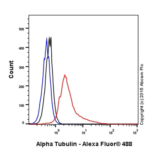
 Flow Cytometry - Anti-alpha Tubulin antibody - Microtubule Marker (ab18251)
Overlay histogram showing Caco2 cells stained with ab18251 (red line). The cells were fixed with 80% methanol (5 min) and then permeabilized with 0.1% PBS-Tween for 20 min. The cells were then incubated in 1x PBS / 10% normal goat serum / 0.3M glycine to block non-specific protein-protein interactions followed by the antibody (ab18251, 0.01μg/1x106 cells) for 30 min at 22°C. The secondary antibody used was Alexa Fluor® 488 goat anti-rabbit IgG (H&L) (ab150081) at 1/4000 dilution for 30 min at 22°C. Isotype control antibody (black line) was rabbit IgG (polyclonal) (ab27478, 0.01μg/1x106 cells) used under the same conditions. Unlabelled sample (blue line) was also used as a control.
Acquisition of >5,000 events were collected using a 20mW Argon ion laser (488nm) and 525/30 bandpass filter.


