Phagocytosis Assay Kit (Red Zymosan) (ab234054)
 (ab234054)")
Key features and details
- Detection method: Fluorescent
- Platform: Microplate reader, Fluor. microscope, Flow cyt.
- Sample type: Adherent cells, Suspension cells
Overview
-
Product name
Phagocytosis Assay Kit (Red Zymosan)
See all Phagocytosis kits -
Detection method
Fluorescent -
Sample type
Adherent cells, Suspension cells -
Species reactivity
Reacts with: Mammals, Other species -
Product overview
Phagocytosis Assay Kit ab234054 uses Zymosan particles which have been pre-labeled with a red dye.
The assay can be used for the rapid and accurate detection and quantification of in vitro phagocytosis by fluorescent microscope, spectrophotometer or flow cytometry. It provides a robust screening system for activators and/or inhibitors of phagocytosis and Toll-like receptors ligands (TLR).
The Zymosan particles used in the phagocytosis assay protocol are labeled for fluorescence at Ex/Em 540/570 nm.
Phagocytosis assay protocol summary:
- add Zymosan to cells and incubate for 2-3 hrs
- harvest cells and resuspend pellet in phagocytosis assay buffer, repeat three times to wash off any remaining Zymosan which is not inside cells
- analyze cells with flow cytometer, fluorescent microscope, or microplate reader -
Notes
In mammals, phagocytosis by phagocytes (e.g., macrophages, dendritic cells, and neutrophils) is essential for a variety of biological events. Phagocytosis comprises a series of events, starting with the binding and recognition of particles by cell surface receptors, followed by the formation of actin-rich membrane extensions around the particle.
Zymosan (Saccharomyces cerevisiae) is prepared from yeast cell wall and consists of protein-carbohydrate complexes. Zymosan is a commonly used pathogen in phagocytosis assays.
-
Platform
Microplate reader, Fluor. microscope, Flow cyt.
Properties
-
Storage instructions
Store at +4°C. Please refer to protocols. -
Components 100 tests Phagocytosis Assay Buffer 2 x 100ml Buffer Additive 2 x 1ml Red Zymosan (Cy3) 1 x 600µl 10X Quenching Solution 1 x 500µl
Images
-
Inhibition of phagocytosis.
J774 macrophages were seeded overnight at 5 x 10⁵ of viable cells/well. The next day the cells were pretreated with 20 µM Cytochalasin D for 1 h at 37ºC prior to addition of 5 µl of Zymosan particles. Phagocytosis was conducted for 2 hours and the amount of engulfed Zymosan was determined as described in the Assay Protocol.
Panel A and B: images of non-treated cells. Panel C and D: treatment with Cytochalasin D.
-
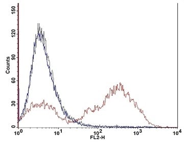 Flow cytometry plot.
J774 macrophages were seeded overnight at 5 x 10⁵ of viable cells/well. The next day the cells were pretreated with 20 µM Cytochalasin D for 1 h at 37ºC prior to addition of 5 µl of Zymosan particles. Phagocytosis was conducted for 2 hours and the amount of engulfed Zymosan was determined as described in the Assay Protocol.
Black line: untreated control cells; red line: macrophages with engulfed Zymosan particles; blue line: inhibition of phagocytosis by Cytochalasin D.
-
 Zymosan Standard curve.
J774 macrophages were seeded overnight at 5 x 10⁵ of viable cells/well. The next day the cells were pretreated with 20 µM Cytochalasin D for 1 h at 37ºC prior to addition of 5 µl of Zymosan particles. Phagocytosis was conducted for 2 hours and the amount of engulfed Zymosan was determined as described in the Assay Protocol.


