Human Peripheral Blood Mononuclear Cell Isolation and Viability Kit (ab234628)
Key features and details
- Sample type: Whole Blood
Overview
-
Product name
Human Peripheral Blood Mononuclear Cell Isolation and Viability Kit -
Sample type
Whole Blood -
Product overview
Human Peripheral Blood Mononuclear Cell Isolation and Viability Kit (ab234628) for PBMC Isolation is the first kit available to provide a complete set of components for the isolation of PBMCs from human blood and also allow visualization of viable cells. High yields of PBMCs (≥2.5 x10⁶ cells/ml) have been obtained following this simple and time-saving protocol. By using the Viability Stain, it was proven that 99% of the cells are viable and the isolated fraction contains low red blood cell counts (≤ 3%).
-
Notes
Peripheral blood mononuclear cells (PBMCs) are found in the peripheral blood circulating throughout the body and not sequestered in the lymphatic system, spleen, liver or bone marrow. PBMCs are composed of four cell types: T cells, B cells, Natural Killer cells and monocytes, each having a round nucleus. Applicable research areas include: stem cell research, evaluation of the immunotherapeutic potential of T or B cells and evaluation of inflammatory response orchestrated by cytokines and chemokines. Based on the unique density of each component, the whole blood is separated into plasma, PBMCs, density gradient media and red blood cells (RBCs).
Properties
-
Storage instructions
Store at +4°C. Please refer to protocols. -
Components 30 ml Blunt-end needle (18 G; 1.5 in., sterile) 1 x 10 units Density Gradient Media 1 x 60ml EDTA (0.5 M, pH 8.0) 1 x 0.5ml Viability Stain 1 x 200µl
Images
-
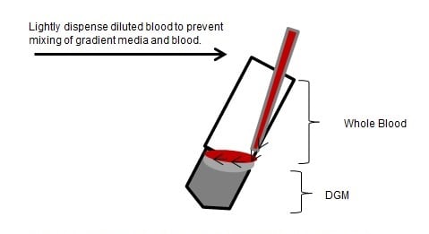
 Illustration of conical tube held at an appropriate angle while blood is layered on top of Density Gradient Media
 -
(1) Layers of Density Gradient Media and whole blood prior to and after centrigugation showing the separation of layers in the conical tube. (2) Illustrates the separation of four layers (plasma, PBMCs, Density Gradient Media and RBCs).
-
Brightfield image of hemocytometer showing total cells (left), image from fluorescent microscope with Rhodamine/FITC filters of the same ROI showing live (green) and dead (red) cells (middle). The two images are merged (right).


