Human NDUFS3 knockout HEK-293T cell line (ab266419)
")
Properties
-
Number of cells
1 x 10⁶ cells/vial, 1 mL -
Viability
~90% -
Adherent /Suspension
Adherent -
Tissue
Kidney -
Cell type
epithelial -
STR Analysis
Amelogenin X D5S818: 8, 9 D13S317: 12, 14 D7S820: 11 D16S539: 9, 13 vWA: 16, 19 TH01: 7, 9.3 TPOX: 11 CSF1PO: 11, 12 -
Antibiotic resistance
Puromycin 1.00µg/ml -
Mycoplasma free
Yes -
Storage instructions
Shipped on Dry Ice. Store in liquid nitrogen. -
Storage buffer
Constituents: 8.7% Dimethylsulfoxide, 2% Cellulose, methyl ether -
Research areas
Images
-
Western blot - Human NDUFS3 knockout HEK293T cell line (ab266419)All lanes : Anti-NDUFS3 antibody [EPR12782] - C-terminal (ab177471) at 1/1000 dilution
Lane 1 : Wild-type HEK-293T cell lysate
Lane 2 : NDUFS3 Knockout HEK-293T cell lysate
Lysates/proteins at 20 µg per lane.
Performed under reducing conditions.
Predicted band size: 30 kDa
Observed band size: 27 kDa why is the actual band size different from the predicted?Lanes 1 - 2: Merged signal (red and green). Green - ab177471 observed at 27 kDa. Red - loading control ab7291 (Mouse anti-Alpha Tubulin [DM1A]) observed at 55kDa.
ab177471 was shown to react with NDUFS3 in HEK-293T wild-type cells in western blot with loss of signal observed in NDUFS3 knockout cell line ab266419 (NDUFS3 knockout cell lysate ab257556). Wild-type and NDUFS3 knockout HEK-293T cell lysates were subjected to SDS-PAGE. Membranes were blocked in 3% milk in TBS-T (0.1% Tween®) before incubation with ab177471 and ab7291 (Mouse anti-Alpha Tubulin [DM1A]) overnight at 4°C at a 1 in 1000 dilution and a 1 in 20000 dilution respectively. Blots were incubated with Goat anti-Rabbit IgG H&L (IRDye® 800CW) preabsorbed (ab216773) and Goat anti-Mouse IgG H&L (IRDye® 680RD) preabsorbed (ab216776) secondary antibodies at 1 in 20000 dilution for 1 hour at room temperature before imaging.
-
 Western blot - Human NDUFS3 knockout HEK293T cell line (ab266419)All lanes : Anti-NDUFS3 antibody [EPR12781] (ab183733) at 1/10000 dilution
Lane 1 : Wild-type HEK-293T cell lysate
Lane 2 : NDUFS3 Knockout HEK-293T cell lysate
Lysates/proteins at 20 µg per lane.
Performed under reducing conditions.
Predicted band size: 30 kDa
Observed band size: 27 kDa why is the actual band size different from the predicted?Lanes 1 - 2: Merged signal (red and green). Green - ab183733 observed at 27 kDa. Red - loading control ab7291 (Mouse anti-Alpha Tubulin [DM1A]) observed at 55kDa.
ab183733 was shown to react with NDUFS3 in HEK-293T wild-type cells in western blot with loss of signal observed in NDUFS3 knockout cell line ab266419 (NDUFS3 knockout cell lysate ab257556). Wild-type and NDUFS3 knockout HEK-293T cell lysates were subjected to SDS-PAGE. Membranes were blocked in 3% milk in TBS-T (0.1% Tween®) before incubation with ab183733 and ab7291 (Mouse anti-Alpha Tubulin [DM1A]) overnight at 4°C at a 1 in 10000 dilution and a 1 in 20000 dilution respectively. Blots were incubated with Goat anti-Rabbit IgG H&L (IRDye® 800CW) preabsorbed (ab216773) and Goat anti-Mouse IgG H&L (IRDye® 680RD) preabsorbed (ab216776) secondary antibodies at 1 in 20000 dilution for 1 hour at room temperature before imaging.
-
 Western blot - Human NDUFS3 knockout HEK293T cell line (ab266419)All lanes : Anti-NDUFS3 antibody [EPR12781] (ab183733) at 1/10000 dilution
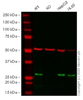
Lane 1 : Wild-type HEK-293T cell lysate
Lane 2 : NDUFS3 knockout HEK-293T cell lysate
Lane 3 : HepG2 cell lysate
Lane 4 : HL60 cell lysate
Lysates/proteins at 20 µg per lane.
Performed under reducing conditions.
Predicted band size: 30 kDa
Observed band size: 30 kDaLanes 1- 4: Merged signal (red and green). Green - ab183733 observed at 30 kDa. Red - Anti-alpha Tubulin antibody [DM1A] - Loading Control (ab7291) observed at 50 kDa.
ab183733 was shown to react with NDUFS3 in wild-type HEK-293T cells in western blot. Loss of signal was observed when knockout cell line ab266419 (knockout cell lysate ab257556) was used. Wild-type HEK-293T and NDUFS3 knockout HEK-293T cell lysates were subjected to SDS-PAGE. Membrane was blocked for 1 hour at room temperature in 0.1% TBST with 3% non-fat dried milk. ab183733 and Anti-alpha Tubulin antibody [DM1A] - Loading Control (ab7291) overnight at 4°C at a 1 in 10000 dilution and a 1 in 20000 dilution respectively. Blots were developed with Goat anti-Rabbit IgG H&L (IRDye®800CW) preadsorbed (ab216773) and Goat anti-Mouse IgG H&L (IRDye®680RD) preadsorbed (ab216776) secondary antibodies at 1 in 20000 dilution for 1 hour at room temperature before imaging.
-
 Western blot - Human NDUFS3 knockout HEK293T cell line (ab266419)All lanes : Anti-NDUFS3 antibody [EPR12782] - C-terminal (ab177471) at 1/1000 dilution
Lane 1 : Wild-type HEK-293T cell lysate
Lane 2 : NDUFS3 knockout HEK-293T cell lysate
Lane 3 : HepG2 cell lysate
Lane 4 : HL60 cell lysate
Lysates/proteins at 20 µg per lane.
Performed under reducing conditions.
Predicted band size: 30 kDa
Observed band size: 30 kDaLanes 1- 4: Merged signal (red and green). Green - ab177471 observed at 30 kDa. Red - Anti-alpha Tubulin antibody [DM1A] - Loading Control (ab7291) observed at 50 kDa.
ab177471 was shown to react with NDUFS3 in wild-type HEK-293T cells in western blot. Loss of signal was observed when knockout cell line ab266419 (knockout cell lysate ab257556) was used. Wild-type HEK-293T and NDUFS3 knockout HEK-293T cell lysates were subjected to SDS-PAGE. Membrane was blocked for 1 hour at room temperature in 0.1% TBST with 3% non-fat dried milk. ab177471 and Anti-alpha Tubulin antibody [DM1A] - Loading Control (ab7291) overnight at 4°C at a 1 in 1000 dilution and a 1 in 20000 dilution respectively. Blots were developed with Goat anti-Rabbit IgG H&L (IRDye®800CW) preadsorbed (ab216773) and Goat anti-Mouse IgG H&L (IRDye®680RD) preadsorbed (ab216776) secondary antibodies at 1 in 20000 dilution for 1 hour at room temperature before imaging.
-
 Sanger Sequencing - Human NDUFS3 knockout HEK293T cell line (ab266419)Homozygous: 19 bp deletion in exon 1


