Human Hif1 + PDK1 Hypoxia In Cell ELISA Kit (IR) (ab125299)
Key features and details
- Assay type: Cell-based (quantitative)
- Detection method: IR
- Sample type: Adherent cells, Suspension cells
- Reacts with: Human
Overview
-
Product name
Human Hif1 + PDK1 Hypoxia In Cell ELISA Kit (IR) -
Detection method
IR -
Precision
Intra-assay Sample n Mean SD CV% HeLa cells -
Sample type
Adherent cells, Suspension cells -
Assay type
Cell-based (quantitative) -
Assay duration
Multiple steps standard assay -
Species reactivity
Reacts with: Human -
Product overview
ab125299 is an In-Cell ELISA (ICE) assay kit that uses quantitative immunocytochemistry to measure HIF1 alpha and PDK1 protein levels in cultured cells. Cells are fixed in a microplate and targets of interest are detected with highly specific, well-characterized antibodies. Relative protein levels are quantified using IRDye®-labeled Secondary Antibodies and IR imaging using a LI-COR® Odyssey® or Aerius® system.
Hypoxia and the cellular response to hypoxic environment is a central topic in studies of metabolism, cancer progression and development and stem cells. A key player is the transcription factor HIF1 alpha (hypoxia inducible factor 1 alpha) which is stabilized at the protein level in response to decreased oxygen tension. HIF1 alpha then promotes transcription of a number of factors that alters cellular physiology. This Hypoxia ICE assay kit provides duplexed measurements of HIF1 alpha and the HIF1 alpha responsive proteins PDK1.
HIF1 alpha (hypoxia-inducible factor 1-alpha) is a constitutively expressed transcription factor that is degraded under normal oxygen tensions but stabilized (at the protein level) when oxygen is limiting (hypoxia). Under hypoxic conditions, stabilized HIF1A promotes the transcription of a host of genes that enable the cell to adapt to the lack of oxygen. A key aspect of the hypoxic response is the switch from aerobic respiration to anaerobic glycolysis and many of the HIF1 alpha responsive genes encode proteins that promote glycolysis and/or inhibit oxidative phosphorylation. Stabilization of the HIF1 alpha protein levels can be detected quickly after the onset of hypoxia, whereas changes in the levels of HIF1 alpha responsive proteins takes longer to manifest. The anti-HIF1 alpha antibody used in this assay is specific for Human HIF1 alpha protein.
PDK1 (dehydrogenase kinase isozyme 1, mitochondrial) is a mitochondrial matrix protein that contributes to the regulation of glucose metabolism. PDK1 inhibits the mitochondrial pyruvate dehydrogenase complex (PDH) by phosphorylation of the E1 alpha subunit. The pyruvate dehydrogenase complex transforms pyruvate to Acetyl-CoA for use in the TCA cycle. Hence, inhibitory phosphorylation events by PDK1 on PDH function to down-regulate TCA and oxidative phosphorylation. Stabilization of the HIF1 alpha transcription factor directly leads to increased transcription of PDK1. The PDK1 antibody used in this assay reacts with Human and Bovine PDK1.
In-Cell ELISA (ICE) technology is used to perform quantitative immunocytochemistry of cultured cells with a near-infrared fluorescent dye-labeled detector antibody. The technique generates quantitative data with specificity similar to Western blotting, but with much greater quantitative precision and higher throughput due to the greater dynamic range and linearity of direct fluorescence detection and the ability to run 96 samples in parallel. This method rapidly fixes the cells in situ, stabilizing the in vivo levels of proteins, and thus essentially eliminates changes during sample handling, such as preparation of protein extracts. Finally, the HIF1 alpha and PDK1 signals can all be normalized to cell amount, measured by the provided Janus Green whole cell stain, to further increase the assay precision.
Plates are available in our ICE (In-Cell ELISA) Support Pack (ab111542) which can be bought seperately.
-
Notes
Upon receipt spin down the contents of the IRDye®-labeled Secondary Antibody tube and protect from light. Store all components upright at 4°C. This kit is stable for at least 6 months from receipt.
Abcam has not and does not intend to apply for the REACH Authorisation of customers’ uses of products that contain European Authorisation list (Annex XIV) substances.
It is the responsibility of our customers to check the necessity of application of REACH Authorisation, and any other relevant authorisations, for their intended uses. -
Platform
Microplate
Properties
-
Storage instructions
Store at +4°C. Please refer to protocols. -
Components 1 x 96 tests 1000X IRDye-labeled Secondary Antibodies 1 x 24µl 100X HIF1alpha Primary Antibody (Rabbit) 1 x 120µl 100X PDK1 Primary Antibody (Mouse) 1 x 120µl 100X Triton X-100 1 x 1.5ml 10X Blocking Solution 1 x 15ml 10X Phosphate Buffered Saline 1 x 100ml 400X Tween-20 1 x 2ml 1X Janus Green Stain 1 x 17ml -
Research areas
-
Cellular localization
Mitochondrial and Nuclear -
Alternative names
- HBP231
- HIF-1
- HIP-1
see all -
Database links
- Entrez Gene: 5163 Human
- Entrez Gene: 29072 Human
- Omim: 602524 Human
- Omim: 612778 Human
- SwissProt: Q9BYW2 Human
- SwissProt: Q15118 Human
Images
-
Coefficient of variation for the experiment described in Figure 1.
-
Antibody specificity demonstrated by western blot. Primary antibodies used in this assay kit were validated by western blot using HeLa cell lysates that had been treated with a dose titration of DFO as indicated. (A) The HIF1 alpha (ab51608) band (indicated by arrow) is absent in untreated cells and induced in a dose-dependent manner by DFO. (B) Similarly, PDK1 levels are increased by DFO treatment in a dose-dependent manner.
-
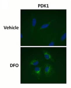
Antibody specificity demonstrated by immunocytochemistry. Primary antibodies used in this assay kit were validated by staining HeLa cells +/- treatment with 1mM DFO (24h) and imaged by fluorescent microscopy. PDK1 staining (ab110335, diluted 2µg/mL) labels cellular mitochondria and the fluorescent intensity increases with DFO treatment.
-
Antibody specificity demonstrated by immunocytochemistry. Primary antibodies used in this assay kit were validated by staining HeLa cells +/- treatment with 1mM DFO (24h) and imaged by fluorescent microscopy. HIF1 alpha (ab51608, diluted 1:2000) staining is absent in untreated cells and induced by DFO treatment. HIF1 alpha localizes to the nucleus (as seen by co-localization with the DNA stain DAPI) as expected.
-
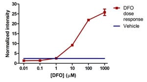
Sample experiment using ab125299 on HeLa cells treated with a titration of DFO. HeLa cells were seeded to an amine coated 96-well microplate and the following day treated with a titration of DFO. After 24h of DFO exposure, the cells were fixed and stained as described in the protocol and the normalized data is presented here +/- SD (as described in the protocol and data analysis sections). PDK1 levels are increased with >10µM DFO.
-
Sample experiment using ab125299 on HeLa cells treated with a titration of DFO. HeLa cells were seeded to an amine coated 96-well microplate and the following day treated with a titration of DFO. After 24h of DFO exposure, the cells were fixed and stained as described in the protocol and the normalized data is presented here +/- SD (as described in the protocol and data analysis sections). HIF1 alpha results show DFO concentrations >10µM induce HIF1A protein levels in a dose dependent fashion.


