Human ERP29 knockout HEK-293T cell line (ab266543)
")
Overview
-
Product name
Human ERP29 knockout HEK-293T cell line -
Description
ERP29 KO HEK-293T cell line -
Parental Cell Line
HEK293T -
Organism
Human -
Mutation description
Knockout achieved by using CRISPR/Cas9, Homozygous: 1 bp insertion in exon 1 -
Passage number
Knockout validation
Sanger Sequencing, Western Blot (WB)Tested applications
Suitable for: WBmore detailsBiosafety level
2General notes
Recommended control: Human wild-type HEK293T cell line (ab255449). Please note a wild-type cell line is not automatically included with a knockout cell line order, if required please add recommended wild-type cell line at no additional cost using the code WILDTYPE-TMTK1.
Cryopreservation cell medium: Cell Freezing Medium-DMSO Serum free media, contains 8.7% DMSO in MEM supplemented with methyl cellulose.
Culture medium: DMEM (High Glucose) + 10% FBS
Initial handling guidelines: Upon arrival, the vial should be stored in liquid nitrogen vapor phase and not at -80ºC. Storage at -80ºC may result in loss of viability.
1. Thaw the vial in 37ºC water bath approximately 1-2 minutes.
2. Transfer the cell suspension (0.8 ml) to a 15 ml/50 ml conical sterile polypropylene centrifuge tube containing 8.4 ml pre-warmed culture medium, wash vial with an additional 0.8 ml culture medium (total volume 10 ml) to collect remaining cells, and centrifuge at 201 x g (rcf) for 5 minutes at room temperature. 10 ml represents minimum recommended dilution. 20 ml represents maximum recommended dilution.
3. Resuspend the cell pellet in 5 ml pre-warmed culture medium and count using a haemocytometer (Click here to view haemocytometer protocol) or alternative cell counting method. Based on cell count, seed cells in an appropriate cell culture flask at a density of 2x10⁴ cells/cm2. This should allow for confluency within 48 hours. Seeding density is given as a guide only and should be scaled to align with individual lab schedules.
4. Incubate the culture at 37ºC incubator with 5% CO2. Cultures should be monitored daily.Subculture guidelines:
- All seeding densities should be based on cell counts gained by established methods.
- A guide seeding density of 2x10⁴ cells/cm2 is recommended for confluency (80-90% confluence) within 48 hours.
- A partial media change 24 hours prior to subculture may be helpful to encourage growth, if required.
- Cells should be passaged when they have achieved 80-90% confluence.
Click here to view the Mammalian cell tissue culture protocol
This product is subject to limited use licenses from The Broad Institute, ERS Genomics Limited and Sigma-Aldrich Co. LLC, and is developed with patented technology. For full details of the licenses and patents please refer to our limited use license and patent pages.
Properties
-
Number of cells
1 x 10⁶ cells/vial, 1 mL -
Viability
~90% -
Adherent /Suspension
Adherent -
Tissue
Kidney -
Cell type
epithelial -
STR Analysis
Amelogenin X D5S818: 8, 9 D13S317: 12, 14 D7S820: 11 D16S539: 9, 13 vWA: 16, 19 TH01: 7, 9.3 TPOX: 11 CSF1PO: 11, 12 -
Antibiotic resistance
Puromycin 1.00µg/ml -
Mycoplasma free
Yes -
Storage instructions
Shipped on Dry Ice. Store in liquid nitrogen. -
Storage buffer
Constituents: 8.7% Dimethylsulfoxide, 2% Cellulose, methyl ether -
Research areas
Target
-
Relevance
Proper protein folding and post-translational modifications are essential for secretory protein export out of the endoplasmic reticulum. This task is accomplished by chaperone proteins such as protein disulfide isomerase (PDI), GRP94, and BiP. A recently characterized protein, designated ERp29, is closely related to these chaperone proteins and appears to be upregulated during ER stress conditions. ERp29 is a soluble 259-residue protein that is localized to the lumen of the endoplasmic reticulum in all mammalian cells. Research has shown that there are two primary domains within ERp29. The first is the C-terminal region that is a novel, all helical, fold that is most likely involved with ERp29 retention to the ER. The second is the N-terminal region that resembles that of PDI’s thioredoxin module. The protein shows sequence similarity to the protein disulfide isomerase family. However, it lacks the thioredoxin motif characteristic of this family, suggesting that this protein does not function as a disulfide isomerase. The protein dimerizes and is thought to play a role in the processing of secretory proteins within the ER. -
Cellular localization
Endoplasmic reticulum, Cell surface.
Properties
-
Number of cells
1 x 10⁶ cells/vial, 1 mL -
Viability
~90% -
Adherent /Suspension
Adherent -
Tissue
Kidney -
Cell type
epithelial -
STR Analysis
Amelogenin X D5S818: 8, 9 D13S317: 12, 14 D7S820: 11 D16S539: 9, 13 vWA: 16, 19 TH01: 7, 9.3 TPOX: 11 CSF1PO: 11, 12 -
Antibiotic resistance
Puromycin 1.00µg/ml -
Mycoplasma free
Yes -
Storage instructions
Shipped on Dry Ice. Store in liquid nitrogen. -
Storage buffer
Constituents: 8.7% Dimethylsulfoxide, 2% Cellulose, methyl ether -
Research areas
Images
-
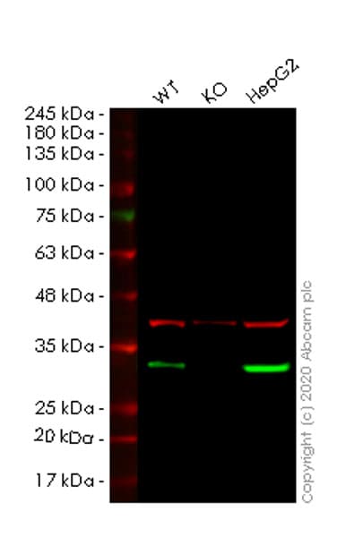
Western blot - Human ERP29 knockout HEK293T cell line (ab266543)All lanes : Anti-ERp29 antibody [EPR12499(B)] (ab176573) at 1/1000 dilution
Lane 1 : Wild-type HEK293T cell lysate
Lane 2 : ERP29 knockout HEK293T cell lysate
Lane 3 : HepG2 cell lysate
Lysates/proteins at 20 µg per lane.
Performed under reducing conditions.
Predicted band size: 29 kDa
Observed band size: 29 kDaLanes 1-3: Merged signal (red and green). Green - ab176573 observed at 29 kDa. Red - loading control, ab8245 observed at 37 kDa.
ab176573 Anti-ERp29 antibody [EPR12499(B)] was shown to specifically react with ERp29 in wild-type HEK293T cells. Loss of signal was observed when knockout cell line ab266543 (knockout cell lysate ab257188) was used. Wild-type and ERp29 knockout samples were subjected to SDS-PAGE. ab176573 and Anti-GAPDH antibody [6C5] - Loading Control (ab8245) were incubated overnight at 4°C at 1 in 1000 dilution and 1 in 20000 dilution respectively. Blots were developed with Goat anti-Rabbit IgG H&L (IRDye® 800CW) preadsorbed (ab216773) and Goat anti-Mouse IgG H&L (IRDye® 680RD) preadsorbed (ab216776) secondary antibodies at 1 in 10000 dilution for 1 hour at room temperature before imaging.
-
 Western blot - Human ERP29 knockout HEK293T cell line (ab266543)All lanes : Anti-ERp29 antibody [EPR12985] (ab175193) at 1/500 dilution
Lane 1 : Wild-type HEK293T cell lysate
Lane 2 : ERP29 knockout HEK293T cell lysate
Lane 3 : HepG2 cell lysate
Lysates/proteins at 20 µg per lane.
Performed under reducing conditions.
Predicted band size: 29 kDa
Observed band size: 29 kDaLanes 1-3: Merged signal (red and green). Green - ab175193 observed at 29 kDa. Red - loading control, ab8245 observed at 37 kDa.
ab175193 Anti-ERp29 antibody [EPR12985] was shown to specifically react with ERp29 in wild-type HEK293T cells. Loss of signal was observed when knockout cell line ab266543 (knockout cell lysate ab257188) was used. Wild-type and ERp29 knockout samples were subjected to SDS-PAGE. ab175193 and Anti-GAPDH antibody [6C5] - Loading Control (ab8245) were incubated overnight at 4°C at 1 in 500 dilution and 1 in 20000 dilution respectively. Blots were developed with Goat anti-Rabbit IgG H&L (IRDye® 800CW) preadsorbed (ab216773) and Goat anti-Mouse IgG H&L (IRDye® 680RD) preadsorbed (ab216776) secondary antibodies at 1 in 10000 dilution for 1 hour at room temperature before imaging.
-
 Sanger Sequencing - Human ERP29 knockout HEK293T cell line (ab266543)Homozygous: 1 bp insertion in exon 1
 -
 Cell Culture - Human ERP29 knockout HEK293T cell line (ab266543)
Representative images of ERP29 knockout HEK293T cells, low and high confluency examples (top left and right respectively) and wild-type HEK293T cells, low and high confluency (bottom left and right respectively) showing typical adherent, epithelial-like morphology. Images were captured at 10X magnification using a EVOS XL Core microscope.