Human CD276 knockout HEK-293T cell line (ab266658)
")
Properties
-
Number of cells
1 x 10⁶ cells/vial, 1 mL -
Viability
~90% -
Adherent /Suspension
Adherent -
Tissue
Kidney -
Cell type
epithelial -
STR Analysis
Amelogenin X D5S818: 8, 9 D13S317: 12, 14 D7S820: 11 D16S539: 9, 13 vWA: 16, 19 TH01: 7, 9.3 TPOX: 11 CSF1PO: 11, 12 -
Mycoplasma free
Yes -
Storage instructions
Shipped on Dry Ice. Store in liquid nitrogen. -
Storage buffer
Constituents: 8.7% Dimethylsulfoxide, 2% Cellulose, methyl ether -
Research areas
Images
-
Western blot - Human CD276 knockout HEK293T cell line (ab266658)All lanes : Anti-CD276 antibody [SP206] (ab227670) at 1/1000 dilution
Lane 2 : CD276 knockout HEK293T cell lysate
Lane 3 : LNCaP cell lysate
Lane 4 : Raji cell lysate
Lysates/proteins at 20 µg per lane.
Secondary
All lanes : Goat anti-Rabbit IgG H&L (IRDye® 800CW) preadsorbed (ab216773) at 1/10000 dilution
Predicted band size: 57 kDa
Observed band size: 90-110 kDa why is the actual band size different from the predicted?Lanes 1-4: Merged signal (red and green). Green - ab227670 observed at 90-110 kDa. Red - loading control ab8245 observed at 36 kDa.
ab227670 Anti-CD276 antibody [SP206] was shown to specifically react with CD276 in wild-type HEK293T cells. Loss of signal was observed when knockout cell line ab266658 (knockout cell lysate ab257097) was used. Wild-type and CD276 knockout samples were subjected to SDS-PAGE. ab227670 and Anti-GAPDH antibody [6C5] - Loading Control (ab8245) were incubated overnight at 4°C at 1 in 1000 dilution and 1 in 20000 dilution respectively. Blots were developed with Goat anti-Rabbit IgG H&L (IRDye® 800CW) preadsorbed (ab216773) and Goat anti-Mouse IgG H&L (IRDye® 680RD) preadsorbed (ab216776) secondary antibodies at 1 in 20000 dilution for 1 hour at room temperature before imaging.
-
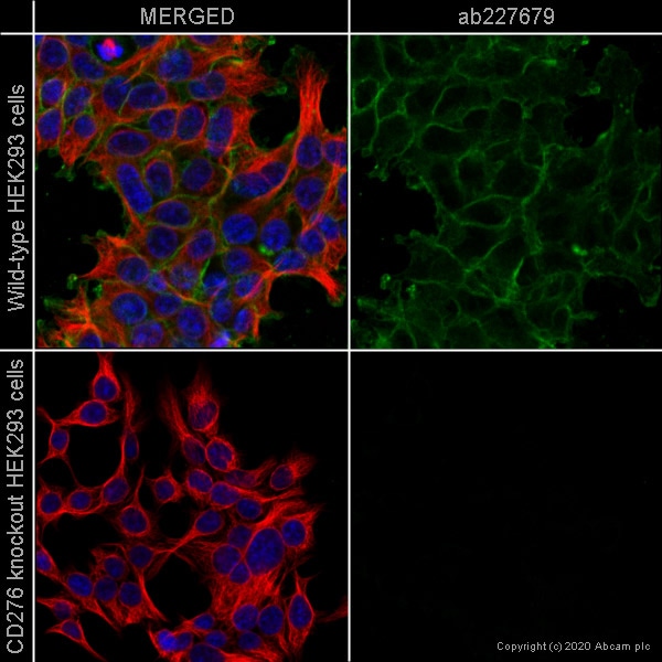 Immunocytochemistry - Human CD276 knockout HEK293T cell line (ab266658)ab227679 staining CD276 in wild-type HEK293 cells (top panel) and CD276 knockout HEK293 cells (ab266658) (bottom panel). The cells were fixed with 100% methanol (5 min) then permeabilized with 0.1% Triton X-100 for 5 minutes and then blocked with 1% BSA/10% normal goat serum/0.3M glycine in 0.1% PBS-Tween for 1h. The cells were then incubated with ab227679 at 1/100 dilution and ab7291 (Mouse monoclonal to alpha Tubulin) at 1/1000 dilution overnight at 4°C followed by a further incubation at room temperature for 1h with a goat secondary antibody to rabbit IgG (Alexa Fluor® 488) (ab150081) at 2 μg/ml (shown in green) and a goat secondary antibody to mouse IgG (Alexa Fluor® 594) (ab150120) at 2 μg/ml (shown in red). Nuclear DNA was labelled in blue with DAPI.
Image was taken with a confocal microscope (Leica-Microsystems TCS SP8). -
 Flow Cytometry - Human CD276 knockout HEK293T cell line (ab266658)
Flow cytometry overlay histogram showing wild-type HEK293 (green line) and CD276 knockout HEK293 cells (ab266658) stained with ab134161 (red line). The cells were incubated in 1x PBS containing 10% normal goat serum to block non-specific protein-protein interaction followed by the antibody (ab134161) (1x10⁶ in 100μl at 0.2 μg/ml) for 30 min at 4°C.
The secondary antibody Goat anti-rabbit IgG H&L (Alexa Fluor® 488, pre-adsorbed) (ab150081) was used at 1/2000 for 30 min at 4°C.
Isotype control antibody was Rabbit IgG (monoclonal) (ab172730) used at the same concentration and conditions as the primary antibody (wild-type HEK293 - black line CD276 HEK293 knockout - grey line). Unlabelled sample was also used as a control (this line is not shown for the purpose of simplicity).
Acquisition of >5000 events were collected using a 50 mW Blue laser (488nm) and 525/40 bandpass filter.
-
 Western blot - Human CD276 knockout HEK293T cell line (ab266658)All lanes : Anti-CD276 antibody [EPR20115] (ab219648) at 1/1000 dilution
Lane 1 : Wild-type HEK293T cell lysate
Lane 2 : CD276 knockout HEK293T cell lysate
Lane 3 : LNCaP cell lysate
Lane 4 : Raji cell lysate
Lysates/proteins at 20 µg per lane.
Secondary
All lanes : Goat anti-Rabbit IgG H&L (IRDye® 800CW) preadsorbed (ab216773) at 1/10000 dilution
Predicted band size: 57 kDa
Observed band size: 90-110 kDa why is the actual band size different from the predicted?Lanes 1-4: Merged signal (red and green). Green - ab219648 observed at 90-110 kDa. Red - loading control ab8245 observed at 36 kDa.
ab219648 Anti-CD276 antibody [EPR20115] was shown to specifically react with CD276 in wild-type HEK293T cells. Loss of signal was observed when knockout cell line ab266658 (knockout cell lysate ab257097) was used. Wild-type and CD276 knockout samples were subjected to SDS-PAGE. ab219648 and Anti-GAPDH antibody [6C5] - Loading Control (ab8245) were incubated overnight at 4°C at 1 in 1000 dilution and 1 in 20000 dilution respectively. Blots were developed with Goat anti-Rabbit IgG H&L (IRDye® 800CW) preadsorbed (ab216773) and Goat anti-Mouse IgG H&L (IRDye® 680RD) preadsorbed (ab216776) secondary antibodies at 1 in 20000 dilution for 1 hour at room temperature before imaging.
-
 Immunocytochemistry - Human CD276 knockout HEK293T cell line (ab266658)ab134161 staining CD276 in wild-type HEK293 cells (top panel) and CD276 knockout HEK293 cells (ab266658) (bottom panel). The cells were fixed with 100% methanol (5 min) then permeabilized with 0.1% Triton X-100 for 5 minutes and then blocked with 1% BSA/10% normal goat serum/0.3M glycine in 0.1% PBS-Tween for 1h. The cells were then incubated with ab134161 at 1μg/ml concentration and ab7291 (Mouse monoclonal to alpha Tubulin) at 1/1000 dilution overnight at 4°C followed by a further incubation at room temperature for 1h with a goat secondary antibody to rabbit IgG (Alexa Fluor® 488) (ab150081) at 2 μg/ml (shown in green) and a goat secondary antibody to mouse IgG (Alexa Fluor® 594) (ab150120) at 2 μg/ml (shown in red). Nuclear DNA was labelled in blue with DAPI.
Image was taken with a confocal microscope (Leica-Microsystems TCS SP8). -
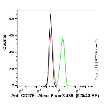 Flow Cytometry - Human CD276 knockout HEK293T cell line (ab266658)
Flow cytometry overlay histogram showing wild-type HEK293 (green line) and CD276 knockout HEK293 cells (ab266658) stained with ab89133 (red line). The cells were incubated in 1x PBS containing 10% normal goat serum to block non-specific protein-protein interaction followed by the antibody (ab89133) (1x10⁶ in 100μl at 0.2 μg/ml) for 30 min at 4°C.
The secondary antibody Goat anti-mouse IgG H&L (Alexa Fluor® 488, pre-adsorbed) (ab150117) was used at 1/2000 for 30 min at 4°C.
Isotype control antibody was mouse IgG1κ (ab170190) used at the same concentration and conditions as the primary antibody (wild-type HEK293 - black line CD276 HEK293 knockout - grey line). Unlabelled sample was also used as a control (this line is not shown for the purpose of simplicity).
Acquisition of >5000 events were collected using a 50 mW Blue laser (488nm) and 525/40 bandpass filter.
-
 Sanger Sequencing - Human CD276 knockout HEK293T cell line (ab266658)Homozygous: Insertion of the selection cassette in exon 2
 -
 Cell Culture - Human CD276 knockout HEK293T cell line (ab266658)
Representative images of CD276 knockout HEK293T cells, low and high confluency examples (top left and right respectively) and wild-type HEK293T cells, low and high confluency (bottom left and right respectively) showing typical adherent, epithelial-like morphology. Images were captured at 10X magnification using a EVOS XL Core microscope.

