Human CANX (Calnexin) knockout HEK-293T cell line (ab255368)
 knockout HEK-293T cell line (ab255368)")
Overview
-
Product name
Human CANX (Calnexin) knockout HEK-293T cell line -
Description
CANX KO HEK-293T cell line -
Parental Cell Line
HEK293T -
Organism
Human -
Mutation description
Knockout achieved by using CRISPR/Cas9, Homozygous: 19 bp deletion in exon 2 -
Passage number
Knockout validation
Sanger Sequencing, Western Blot (WB)Tested applications
Suitable for: WBmore detailsBiosafety level
2General notes
Recommended control: Human wild-type HEK293T cell line (ab255449). Please note a wild-type cell line is not automatically included with a knockout cell line order, if required please add recommended wild-type cell line at no additional cost using the code WILDTYPE-TMTK1.
Cryopreservation cell medium: Cell Freezing Medium-DMSO Serum free media, contains 8.7% DMSO in MEM supplemented with methyl cellulose.
Culture medium: DMEM (High Glucose) + 10% FBS
Initial handling guidelines: Upon arrival, the vial should be stored in liquid nitrogen vapor phase and not at -80ºC. Storage at -80ºC may result in loss of viability.
1. Thaw the vial in 37ºC water bath approximately 1-2 minutes.
2. Transfer the cell suspension (0.8 ml) to a 15 ml/50 ml conical sterile polypropylene centrifuge tube containing 8.4 ml pre-warmed culture medium, wash vial with an additional 0.8 ml culture medium (total volume 10 ml) to collect remaining cells, and centrifuge at 201 x g (rcf) for 5 minutes at room temperature. 10 ml represents minimum recommended dilution. 20 ml represents maximum recommended dilution.
3. Resuspend the cell pellet in 5 ml pre-warmed culture medium and count using a haemocytometer (Click here to view haemocytometer protocol) or alternative cell counting method. Based on cell count, seed cells in an appropriate cell culture flask at a density of 2x10⁴ cells/cm2. This should allow for confluency within 48 hours. Seeding density is given as a guide only and should be scaled to align with individual lab schedules.
4. Incubate the culture at 37ºC incubator with 5% CO2. Cultures should be monitored daily.Subculture guidelines:
- All seeding densities should be based on cell counts gained by established methods.
- A guide seeding density of 2x10⁴ cells/cm2 is recommended for confluency (80-90% confluence) within 48 hours.
- A partial media change 24 hours prior to subculture may be helpful to encourage growth, if required.
- Cells should be passaged when they have achieved 80-90% confluence.
Click here to view the Mammalian cell tissue culture protocol
This product is subject to limited use licenses from The Broad Institute and ERS Genomics Limited, and is developed with patented technology. For full details of the limited use licenses and relevant patents please refer to our limited use license and patent pages.
Properties
-
Number of cells
1 x 10⁶ cells/vial, 1 mL -
Viability
~90% -
Adherent /Suspension
Adherent -
Tissue
Kidney -
Cell type
epithelial -
STR Analysis
Amelogenin X D5S818: 8, 9 D13S317: 12, 14 D7S820: 11 D16S539: 9, 13 vWA: 16, 19 TH01: 7, 9.3 TPOX: 11 CSF1PO: 11, 12 -
Mycoplasma free
Yes -
Storage instructions
Shipped on Dry Ice. Store in liquid nitrogen. -
Storage buffer
Constituents: 8.7% Dimethylsulfoxide, 2% Cellulose, methyl ether -
Research areas
Target
-
Function
Calcium-binding protein that interacts with newly synthesized glycoproteins in the endoplasmic reticulum. It may act in assisting protein assembly and/or in the retention within the ER of unassembled protein subunits. It seems to play a major role in the quality control apparatus of the ER by the retention of incorrectly folded proteins. -
Sequence similarities
Belongs to the calreticulin family. -
Cellular localization
Endoplasmic reticulum membrane. Melanosome. Identified by mass spectrometry in melanosome fractions from stage I to stage IV. - Information by UniProt
Properties
-
Number of cells
1 x 10⁶ cells/vial, 1 mL -
Viability
~90% -
Adherent /Suspension
Adherent -
Tissue
Kidney -
Cell type
epithelial -
STR Analysis
Amelogenin X D5S818: 8, 9 D13S317: 12, 14 D7S820: 11 D16S539: 9, 13 vWA: 16, 19 TH01: 7, 9.3 TPOX: 11 CSF1PO: 11, 12 -
Mycoplasma free
Yes -
Storage instructions
Shipped on Dry Ice. Store in liquid nitrogen. -
Storage buffer
Constituents: 8.7% Dimethylsulfoxide, 2% Cellulose, methyl ether -
Research areas
Images
-
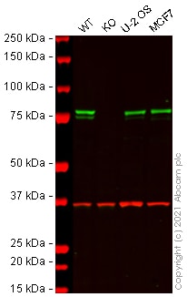
Western blot - Human CANX (Calnexin) knockout HEK-293T cell line (ab255368)All lanes : Anti-Calnexin antibody [EPR3632] (ab92573) at 1/20000 dilution
Lane 1 : Wild-type HEK-293T cell lysate
Lane 2 : CANX knockout HEK-293T cell lysate
Lane 3 : U-2 OS cell lysate
Lane 4 : MCF7 cell lysate
Lysates/proteins at 20 µg per lane.
Performed under reducing conditions.
Predicted band size: 68 kDa
Observed band size: 80 kDa why is the actual band size different from the predicted?Lanes 1 - 4: Merged signal (red and green). Green - ab92573 observed at 80 kDa. Red - loading control ab8245 (Mouse anti-GAPDH antibody [6C5]) observed at 37 kDa.
ab92573 was shown to react with Calnexin in wild-type HEK-293T cells in Western blot with loss of signal observed in CANX knockout cell line ab255368 (CANX knockout cell lysate ab263805). Wild-type HEK-293T and CANX knockout cell lysates were subjected to SDS-PAGE. Membranes were blocked in 3 % milk in TBS-T (0.1 % Tween®) before incubation with ab92573 and ab8245 (Mouse anti-GAPDH antibody [6C5]) overnight at 4 °C at a 1 in 20000 dilution and a 1 in 20000 dilution respectively. Blots were incubated with Goat anti-Rabbit IgG H&L (IRDye® 800CW) preabsorbed (ab216773) and Goat anti-Mouse IgG H&L (IRDye® 680RD) preabsorbed (ab216776) secondary antibodies at 1 in 20000 dilution for 1 h at room temperature before imaging.
-
 Western blot - Human CANX (Calnexin) knockout HEK293T cell line (ab255368)All lanes : Anti-Calnexin antibody [CANX/1543] (ab238078) at 1 µg/ml
Lane 1 : Wild-type HEK-293T cell lysate
Lane 2 : CANX knockout HEK-293T cell lysate
Lane 3 : U-2 OS cell lysate
Lane 4 : MCF7 cell lysate
Lysates/proteins at 20 µg per lane.
Performed under reducing conditions.
Predicted band size: 68 kDa
Observed band size: 80 kDa why is the actual band size different from the predicted?Lanes 1 - 4: Merged signal (red and green). Green - ab238078 observed at 80 kDa. Red - loading control ab181602 (Rabbit Anti-GAPDH antibody [EPR16891]) observed at 37 kDa.
ab238078 was shown to react with Calnexin in wild-type HEK-293T cells in Western blot with loss of signal observed in CANX knockout cell line ab255368 (CANX knockout cell lysate ab263805). Wild-type HEK-293T and CANX knockout cell lysates were subjected to SDS-PAGE. Membranes were blocked in 3 % milk in TBS-T (0.1 % Tween®) before incubation with ab238078 and ab181602 (Rabbit Anti-GAPDH antibody [EPR16891]) overnight at 4 °C at 1 µg/ml and a 1 in 20000 dilution respectively. Blots were incubated with Goat anti-Mouse IgG H&L (IRDye® 800CW) preabsorbed (ab216772) and Goat anti-Rabbit IgG H&L (IRDye® 680RD) preabsorbed (ab216777) secondary antibodies at 1 in 20000 dilution for 1 h at room temperature before imaging.
-
 Western blot - Human CANX (Calnexin) knockout HEK293T cell line (ab255368)All lanes : Anti-Calnexin antibody [EPR3633(2)] - ER Membrane Marker (ab133615) at 1/1000 dilution
Lane 1 : Wild-type HEK-293T cell lysate
Lane 2 : CANX knockout HEK-293T cell lysate
Lysates/proteins at 20 µg per lane.
Performed under reducing conditions.
Predicted band size: 68 kDa
Observed band size: 90 kDa why is the actual band size different from the predicted?Lanes 1- 2: Merged signal (red and green). Green - ab133615 observed at 90 kDa. Red - Anti-GAPDH antibody [6C5] - Loading Control (ab8245) observed at 37 kDa.
ab133615 was shown to react with CANX in wild-type HEK-293T cells in western blot. Loss of signal was observed when knockout cell line ab255368 (knockout cell lysate ab263805) was used. Wild-type HEK-293T and CANX knockout HEK-293T cell lysates were subjected to SDS-PAGE. Membrane was blocked for 1 hour at room temperature in 0.1% TBST with 3% non-fat dried milk. ab133615 and Anti-GAPDH antibody [6C5] - Loading Control (ab8245) overnight at 4°C at a 1 in 1000 dilution and a 1 in 20000 dilution respectively. Blots were developed with Goat anti-Rabbit IgG H&L (IRDye®800CW) preadsorbed (ab216773) and Goat anti-Mouse IgG H&L (IRDye®680RD) preadsorbed (ab216776) secondary antibodies at 1 in 20000 dilution for 1 hour at room temperature before imaging.
-
 Sanger Sequencing - Human CANX knockout HEK293T cell line (ab255368)Homozygous: 19 bp deletion in exon2


