Human BSG (CD147) knockout A549 cell line (ab273748)
 knockout A549 cell line (ab273748)")
Properties
-
Number of cells
1 x 10⁶ cells/vial, 1 mL -
Viability
~90% -
Adherent /Suspension
Adherent -
Tissue
Lung -
Cell type
epithelial -
Disease
Carcinoma -
Gender
Male -
Mycoplasma free
Yes -
Storage instructions
Shipped on Dry Ice. Store in liquid nitrogen. -
Storage buffer
Constituents: 8.7% Dimethylsulfoxide, 2% Cellulose, methyl ether -
Purity
Immunogen affinity purified -
Research areas
Images
-
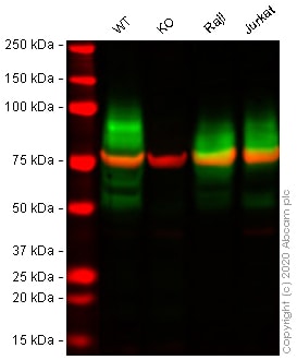
Western blot - Human BSG (CD147) knockout A549 cell line (ab273748)All lanes : Anti-CD147 antibody [MEM-M6/1] (ab666) at 1 µg/ml
Lane 1 : Wild-type A549 cell lysate
Lane 2 : BSG knockout A549 cell lysate
Lane 3 : Raji cell lysate
Lane 4 : Jurkat cell lysate
Lysates/proteins at 30 µg per lane.
Performed under reducing conditions.
Predicted band size: 42 kDa
Observed band size: 55-70 kDa why is the actual band size different from the predicted?Lanes 1 - 4: Merged signal (red and green). Green - ab666 observed at 55-70 kDa. Red - loading control ab52866 (Rabbit anti-alpha Tubulin antibody [EP1332Y]) observed at 55kDa.
ab666 was shown to react with CD147 in wild-type A549 cells in western blot with loss of signal observed in BSG knockout cell line ab273748 (knockout cell lysate ab275500). Wild-type and BSG knockout A549 cell lysates were subjected to SDS-PAGE. Membranes were blocked in fluorescent western blot (TBS-based) blocking solution before incubation with ab666 and ab52866 (Rabbit anti-alpha Tubulin antibody [EP1332Y]) overnight at 4°C at 1 µg/ml and a 1 in 20000 dilution respectively. Blots were incubated with Goat anti-Mouse IgG H&L (IRDye® 800CW) preabsorbed (ab216772) and Goat anti-Rabbit IgG H&L (IRDye® 680RD) preabsorbed (ab216777) secondary antibodies at 1 in 20000 dilution for 1 hour at room temperature before imaging.
-
 Flow Cytometry - Human BSG (CD147) knockout A549 cell line (ab273748)
Flow cytometry overlay histogram showing wild-type A549 (green line) and BSG knockout A549 cells (ab273748) stained with ab666 (red line). The cells were incubated in 1x PBS containing 10% normal goat serum to block non-specific protein-protein interaction followed by the antibody (ab666) (1x10⁶ in 100μl at 10 μg/ml) for 30 min at 4°C.
The secondary antibody Goat anti-mouse IgG H&L (Alexa Fluor® 488, pre-adsorbed) (ab150117) was used at 1/2000 for 30 min at 4°C.
Isotype control antibody was mouse IgG1κ (ab170190) used at the same concentration and conditions as the primary antibody (wild-type A549 - black line; BSG knockout A549 - grey line). Unlabelled sample was also used as a control (this line is not shown for the purpose of simplicity).
Acquisition of >5000 events were collected using a 50 mW Blue laser (488nm) and 525/40 bandpass filter.
-
 Sandwich ELISA - Human BSG (CD147) knockout A549 cell line (ab273748)Human CD147 concentration was interpolated from the EMMPRIN (CD147) standard curve. Supernatants from cell culture samples were serially diluted and assessed by the Human EMMPRIN ELISA kit (ab219631). Wild-type and CD147 knockout A549 cells (ab273748) were assessed in duplicate (n=2); Jurkat and Raji cells were used as positive and negative controls respectively (n=1). Where samples were run in duplicate, data are represented as the mean and error bars represent standard deviation.
 -
 Western blot - Human BSG (CD147) knockout A549 cell line (ab273748)All lanes : Anti-CD147 antibody [EPR4053] (ab108308) at 1/1000 dilution
Lane 1 : Wild-type A549 cell lysate
Lane 2 : BSG knockout A549 cell lysate
Lane 3 : Raji cell lysate
Lysates/proteins at 30 µg per lane.
Performed under reducing conditions.
Predicted band size: 42 kDa
Observed band size: 42-70 kDa why is the actual band size different from the predicted?Lanes 1 - 3: Merged signal (red and green). Green - ab108308 observed at 42-70 kDa. Red - loading control ab7291 (Mouse anti-Alpha Tubulin [DM1A] observed at 55kDa.
ab108308 was shown to react with CD147 in wild-type A549 cells in western blot with loss of signal observed in BSG knockout cell line ab273748 (knockout cell lysate ab275500). Wild-type and BSG knockout A549 cell lysates were subjected to SDS-PAGE. Membranes were blocked in fluorescent western blot (TBS-based) blocking solution before incubation with ab108308 and ab7291 (Mouse anti-Alpha Tubulin [DM1A] overnight at 4°C at a 1 in 1000 Dilution and a 1 in 20000 dilution respectively. Blots were incubated with Goat anti-Rabbit IgG H&L (IRDye® 800CW) preabsorbed (ab216773) and Goat anti-Mouse IgG H&L (IRDye® 680RD) preabsorbed (ab216776) secondary antibodies at 1 in 20000 dilution for 1 hour at room temperature before imaging.
-
 Western blot - Human BSG (CD147) knockout A549 cell line (ab273748)All lanes : Anti-CD147 antibody [OTI9B10] (ab119020) at 1/2000 dilution
Lane 1 : Wild-type A549 cell lysate
Lane 2 : BSG knockout A549 cell lysate
Lane 3 : Raji cell lysate
Lane 4 : Jurkat cell lysate
Lysates/proteins at 30 µg per lane.
Performed under reducing conditions.
Predicted band size: 42 kDa
Observed band size: 55-70 kDa why is the actual band size different from the predicted?Lanes 1 - 4: Merged signal (red and green). Green - ab119020 observed at 55-70 kDa. Red - loading control ab52866 (Rabbit anti-alpha Tubulin antibody [EP1332Y]) observed at 55kDa.
ab119020 was shown to react with CD147 in wild-type A549 cells in western blot with loss of signal observed in BSG knockout cell line ab273748 (knockout cell lysate ab275500). Wild-type and BSG knockout A549 cell lysates were subjected to SDS-PAGE. Membranes were blocked in fluorescent western blot (TBS-based) blocking solution before incubation with ab119020 and ab52866 (Rabbit anti-alpha Tubulin antibody [EP1332Y]) overnight at 4°C at a 1 in 2000 Dilution and a 1 in 20000 dilution respectively. Blots were incubated with Goat anti-Mouse IgG H&L (IRDye® 800CW) preabsorbed (ab216772) and Goat anti-Rabbit IgG H&L (IRDye® 680RD) preabsorbed (ab216777) secondary antibodies at 1 in 20000 dilution for 1 hour at room temperature before imaging.
-
 Western blot - Human BSG (CD147) knockout A549 cell line (ab273748)All lanes : Anti-CD147 antibody [10E10] (ab230921) at 1/500 dilution
Lane 1 : Wild-type A549 cell lysate
Lane 2 : BSG knockout A549 cell lysate
Lane 3 : Raji cell lysate
Lane 4 : Jurkat cell lysate
Lysates/proteins at 30 µg per lane.
Performed under reducing conditions.
Predicted band size: 42 kDa
Observed band size: 42-70 kDa why is the actual band size different from the predicted?Lanes 1 - 4: Merged signal (red and green). Green - ab230921 observed at 42-70 kDa. Red - loading control ab52866 (Rabbit anti-alpha Tubulin antibody [EP1332Y]) observed at 55kDa.
ab230921 was shown to react with CD147 in wild-type A549 cells in western blot with loss of signal observed in BSG knockout cell line ab273748 (knockout cell lysate ab275500). Wild-type and BSG knockout A549 cell lysates were subjected to SDS-PAGE. Membranes were blocked in fluorescent western blot (TBS-based) blocking solution before incubation with ab230921 and ab52866 (Rabbit anti-alpha Tubulin antibody [EP1332Y]) overnight at 4°C at a 1 in 500 Dilution and a 1 in 20000 dilution respectively. Blots were incubated with Goat anti-Mouse IgG H&L (IRDye® 800CW) preabsorbed (ab216772) and Goat anti-Rabbit IgG H&L (IRDye® 680RD) preabsorbed (ab216777) secondary antibodies at 1 in 20000 dilution for 1 hour at room temperature before imaging.
-
 Flow Cytometry - Human BSG (CD147) knockout A549 cell line (ab273748)
Flow cytometry overlay histogram showing wild-type A549 (green line) and BSG knockout A549 cells (ab273748) stained with ab194401 (red line). The cells were incubated in 1x PBS containing 10% normal goat serum to block non-specific protein-protein interaction followed by the antibody (ab194401) (1x10⁶ in 100μl at 10 μg/ml) for 30 min at 4°C.
The secondary antibody Goat anti-mouse IgG H&L (Alexa Fluor® 488, pre-adsorbed) (ab150117) was used at 1/2000 for 30 min at 4°C.
Isotype control antibody was mouse IgG1κ (ab170190) used at the same concentration and conditions as the primary antibody (wild-type A549 - black line; BSG knockout A549 - grey line). Unlabelled sample was also used as a control (this line is not shown for the purpose of simplicity).
Acquisition of >5000 events were collected using a 50 mW Blue laser (488nm) and 525/40 bandpass filter.
-
 Flow Cytometry - Human BSG (CD147) knockout A549 cell line (ab273748)
Flow cytometry overlay histogram showing wild-type A549 (green line) and BSG knockout A549 cells (ab273748) stained with ab91147 (red line). The cells were incubated in 1x PBS containing 10% normal goat serum to block non-specific protein-protein interaction followed by the antibody (ab91147) (1x10⁶ in 100μl at 10 μg/ml) for 30 min at 4°C.
The secondary antibody Goat anti-mouse IgG H&L (Alexa Fluor® 488, pre-adsorbed) (ab150117) was used at 1/2000 for 30 min at 4°C.
Isotype control antibody was mouse IgG1κ (ab170190) used at the same concentration and conditions as the primary antibody (wild-type A549 - black line; BSG knockout A549 - grey line). Unlabelled sample was also used as a control (this line is not shown for the purpose of simplicity).
Acquisition of >5000 events were collected using a 50 mW Blue laser (488nm) and 525/40 bandpass filter.
-
 Flow Cytometry - Human BSG (CD147) knockout A549 cell line (ab273748)
Flow cytometry overlay histogram showing wild-type A549 (green line) and BSG knockout A549 cells (ab273748) stained with ab21903 (red line). The cells were incubated in 1x PBS containing 10% normal goat serum to block non-specific protein-protein interaction followed by the antibody (ab21903) (1x10⁶ in 100μl at 5 μg/ml) for 30 min at 4°C.
The secondary antibody Goat anti-mouse IgG H&L (Alexa Fluor® 488, pre-adsorbed) (ab150117) was used at 1/2000 for 30 min at 4°C.
Isotype control antibody was mouse IgG2bκ (ab170192) used at the same concentration and conditions as the primary antibody (wild-type A549 - black line; BSG knockout A549 - grey line). Unlabelled sample was also used as a control (this line is not shown for the purpose of simplicity).
Acquisition of >5000 events were collected using a 50 mW Blue laser (488nm) and 525/40 bandpass filter.
-
 Flow Cytometry - Human BSG (CD147) knockout A549 cell line (ab273748)
Flow cytometry overlay histogram showing wild-type A549 (green line) and BSG knockout A549 cells (ab273748) stained with ab230921 (red line). The cells were incubated in 1x PBS containing 10% normal goat serum to block non-specific protein-protein interaction followed by the antibody (ab230921) (1x10⁶ in 100μl at 5 μg/ml) for 30 min at 4°C.
The secondary antibody Goat anti-mouse IgG H&L (Alexa Fluor® 488, pre-adsorbed) (ab150117) was used at 1/2000 for 30 min at 4°C.
Isotype control antibody was mouse IgG1κ (ab170190) used at the same concentration and conditions as the primary antibody (wild-type A549 - black line; BSG knockout A549 - grey line). Unlabelled sample was also used as a control (this line is not shown for the purpose of simplicity).
Acquisition of >5000 events were collected using a 50 mW Blue laser (488nm) and 525/40 bandpass filter.
-
 Sanger Sequencing - Human BSG (CD147) knockout A549 cell line (ab273748)
Allele-1: 78 bp insertion in exon 5 introducing premature STOP codon.


