Human BAK1 (Bak) knockout HeLa cell line (ab265277)
 knockout HeLa cell line (ab265277)")
Overview
-
Product name
Human BAK1 (Bak) knockout HeLa cell line
See all Bak lysates -
Parental Cell Line
HeLa -
Organism
Human -
Mutation description
Knockout achieved by using CRISPR/Cas9, 1 bp deletion in exon 2 and 2 bp deletion in exon 2 and Insertion of the selection cassette in exon 2 -
Passage number
Knockout validation
Sanger Sequencing, Western Blot (WB)Tested applications
Suitable for: WBmore detailsBiosafety level
2General notes
Recommended control: Human wild-type HeLa cell line (ab255928). Please note a wild-type cell line is not automatically included with a knockout cell line order, if required please add recommended wild-type cell line at no additional cost using the code WILDTYPE-TMTK1.
Cryopreservation cell medium: Cell Freezing Medium-DMSO Serum free media, contains 8.7% DMSO in MEM supplemented with methyl cellulose.
Culture medium: DMEM (High Glucose) + 10% FBS
Initial handling guidelines: Upon arrival, the vial should be stored in liquid nitrogen vapor phase and not at -80ºC. Storage at -80ºC may result in loss of viability.
1. Thaw the vial in 37ºC water bath approximately 1-2 minutes.
2. Transfer the cell suspension (0.8 ml) to a 15 ml/50 ml conical sterile polypropylene centrifuge tube containing 8.4 ml pre-warmed culture medium, wash vial with an additional 0.8 ml culture medium (total volume 10 ml) to collect remaining cells, and centrifuge at 201 x g (rcf) for 5 minutes at room temperature. 10 ml represents minimum recommended dilution. 20 ml represents maximum recommended dilution.
3. Resuspend the cell pellet in 5 ml pre-warmed culture medium and count using a haemocytometer (Click here to view haemocytometer protocol) or alternative cell counting method. Based on cell count, seed cells in an appropriate cell culture flask at a density of 2x10⁴ cells/cm2. This should allow for confluency within 48 hours. Seeding density is given as a guide only and should be scaled to align with individual lab schedules.
4. Incubate the culture at 37ºC incubator with 5% CO2. Cultures should be monitored daily.Subculture guidelines:
- All seeding densities should be based on cell counts gained by established methods.
- A guide seeding density of 2x10⁴ cells/cm2 is recommended for confluency (80-90% confluence) within 48 hours.
- A partial media change 24 hours prior to subculture may be helpful to encourage growth, if required.
- Cells should be passaged when they have achieved 80-90% confluence.
Click here to view the Mammalian cell tissue culture protocol
This product is subject to limited use licenses from The Broad Institute, ERS Genomics Limited and Sigma-Aldrich Co. LLC, and is developed with patented technology. For full details of the licenses and patents please refer to our limited use license and patent pages.
Properties
-
Number of cells
1 x 10⁶ cells/vial, 1 mL -
Viability
~90% -
Adherent /Suspension
Adherent -
Tissue
Cervix -
Cell type
epithelial -
Disease
Adenocarcinoma -
Gender
Female -
STR Analysis
Amelogenin X D5S818: 11, 12 D13S317: 12, 13.3 D7S820: 8, 12 D16S539: 9, 10 vWA: 16, 18 TH01: 7 TPOX: 8,12 CSF1PO: 9, 10 -
Mycoplasma free
Yes -
Storage instructions
Shipped on Dry Ice. Store in liquid nitrogen. -
Storage buffer
Constituents: 8.7% Dimethylsulfoxide, 2% Cellulose, methyl ether -
Research areas
Target
-
Function
In the presence of an appropriate stimulus, accelerates programmed cell death by binding to, and antagonizing the anti-apoptotic action of BCL2 or its adenovirus homolog E1B 19k protein. Low micromolar levels of zinc ions inhibit the promotion of apoptosis. -
Tissue specificity
Expressed in a wide variety of tissues, with highest levels in the heart and skeletal muscle. -
Sequence similarities
Belongs to the Bcl-2 family. -
Domain
Intact BH3 motif is required by BIK, BID, BAK, BAD and BAX for their pro-apoptotic activity and for their interaction with anti-apoptotic members of the Bcl-2 family. -
Cellular localization
Mitochondrion membrane. - Information by UniProt
Properties
-
Number of cells
1 x 10⁶ cells/vial, 1 mL -
Viability
~90% -
Adherent /Suspension
Adherent -
Tissue
Cervix -
Cell type
epithelial -
Disease
Adenocarcinoma -
Gender
Female -
STR Analysis
Amelogenin X D5S818: 11, 12 D13S317: 12, 13.3 D7S820: 8, 12 D16S539: 9, 10 vWA: 16, 18 TH01: 7 TPOX: 8,12 CSF1PO: 9, 10 -
Mycoplasma free
Yes -
Storage instructions
Shipped on Dry Ice. Store in liquid nitrogen. -
Storage buffer
Constituents: 8.7% Dimethylsulfoxide, 2% Cellulose, methyl ether -
Research areas
Images
-
Western blot - Human BAK1 (Bak) knockout HeLa cell line (ab265277)All lanes : Anti-Bak antibody (ab92999)
Lane 1 : Wild-type HeLa cell lysate
Lane 2 : BAK1 knockout HeLa cell lysate
Lysates/proteins at 20 µg per lane.
Performed under reducing conditions.
Predicted band size: 23 kDa
Observed band size: 23 kDaLanes 1- 2: Merged signal (red and green). Green - ab92999 observed at 23 kDa. Red - Anti-GAPDH antibody [6C5] - Loading Control (ab8245) observed at 37 kDa.
ab92999 was shown to react with Bak in wild-type HeLa cells in western blot. Loss of signal was observed when knockout cell line ab265277 (knockout cell lysate ab257077) was used. Wild-type HeLa and BAK1 knockout HeLa cell lysates were subjected to SDS-PAGE. Membrane was blocked for 1 hour at room temperature in 0.1% TBST with 3% non-fat dried milk. ab92999 and Anti-GAPDH antibody [6C5] - Loading Control (ab8245) overnight at 4°C at 1 µg/ml and a 1 in 20000 dilution respectively. Blots were developed with Goat anti-Rabbit IgG H&L (IRDye®800CW) preadsorbed (ab216773) and Goat anti-Mouse IgG H&L (IRDye®680RD) preadsorbed (ab216776) secondary antibodies at 1 in 20000 dilution for 1 hour at room temperature before imaging.
-
 Western blot - Human BAK1 (Bak) knockout HeLa cell line (ab265277)All lanes : Anti-Bak antibody [Y164] (ab32371) at 1/1000 dilution
Lane 1 : Wild-type HeLa cell lysate
Lane 2 : BAK1 knockout HeLa cell lysate
Lysates/proteins at 20 µg per lane.
Performed under reducing conditions.
Predicted band size: 23 kDa
Observed band size: 23 kDaLanes 1- 2: Merged signal (red and green). Green - ab32371 observed at 23 kDa. Red - Anti-GAPDH antibody [6C5] - Loading Control (ab8245) observed at 37 kDa.
ab32371 was shown to react with Bak in wild-type HeLa cells in western blot. Loss of signal was observed when knockout cell line ab265277 (knockout cell lysate ab257077) was used. Wild-type HeLa and BAK1 knockout HeLa cell lysates were subjected to SDS-PAGE. Membrane was blocked for 1 hour at room temperature in 0.1% TBST with 3% non-fat dried milk. ab32371 and Anti-GAPDH antibody [6C5] - Loading Control (ab8245) overnight at 4°C at a 1 in 1000 dilution and a 1 in 20000 dilution respectively. Blots were developed with Goat anti-Rabbit IgG H&L (IRDye®800CW) preadsorbed (ab216773) and Goat anti-Mouse IgG H&L (IRDye®680RD) preadsorbed (ab216776) secondary antibodies at 1 in 20000 dilution for 1 hour at room temperature before imaging.
-
 Sanger Sequencing - Human BAK1 knockout HeLa cell line (ab265277)
Allele-1: 2 bp deletion in exon 2.
-
 Sanger Sequencing - Human BAK1 knockout HeLa cell line (ab265277)
Allele-2: 1 bp deletion in exon 2.
-
 Sanger Sequencing - Human BAK1 knockout HeLa cell line (ab265277)
Allele-3: Insertion of the selection cassette in exon 2.
-
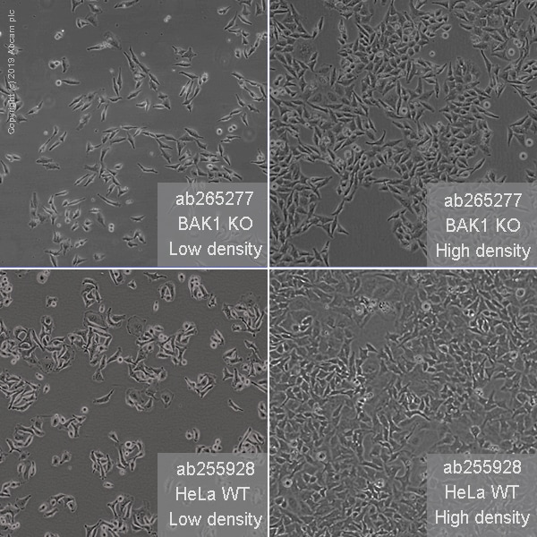 Cell Culture - Human BAK1 (Bak) knockout HeLa cell line (ab265277)
Representative images of BAK1 knockout HeLa cells, low and high confluency examples (top left and right respectively) and wild-type HeLa cells, low and high confluency (bottom left and right respectively) showing typical adherent, epithelial-like morphology. Images were captured at 10X magnification using a EVOS XL Core microscope.


