Goat Anti-Rabbit IgG H&L (HRP) (ab97051)
Key features and details
- Goat Anti-Rabbit IgG H&L (HRP)
- Conjugation: HRP
- Host species: Goat
- Isotype: IgG
- Suitable for: ICC, IHC-P, ELISA, WB
Overview
-
Product name
Goat Anti-Rabbit IgG H&L (HRP)
See all IgG secondary antibodies -
Host species
Goat -
Target species
Rabbit -
Specificity
By immunoelectrophoresis and ELISA this antibody reacts specifically with Rabbit IgG and with light chains common to other Rabbit immunoglobulins. No antibody was detected against non-immunoglobulin serum proteins. -
Tested applications
Suitable for: ICC, IHC-P, ELISA, WBmore details -
Conjugation
HRP
Properties
-
Form
Liquid -
Storage instructions
Shipped at 4°C. Store at +4°C. -
Storage buffer
pH: 6.8
Constituents: 0.2% BSA, PBS, 0.05% CMIT/MIT based preservative -
 Concentration information loading...
 -
Purity
Immunogen affinity purified -
Purification notes
This antibody was isolated by affinity chromatography using antigen coupled to agarose beads and conjugated to Horse Radish Peroxidase (HRP). -
Conjugation notes
Molar enzyme/ antibody protein ratio is 4:1 -
Clonality
Polyclonal -
Isotype
IgG -
General notes
Part of the AbExcel range. -
Research areas
Images
-
All lanes : Anti-Estrogen Receptor alpha antibody [E115] - ChIP Grade (ab32063) at 1/1000 dilution
Lane 1 : Rat pituitary whole tissue lysate
Lane 2 : Mouse pituitary whole tissue lysate
Lysates/proteins at 20 µg per lane.
Secondary
All lanes : Goat Anti-Rabbit IgG H&L (HRP) (ab97051) at 1/20000 dilutionExposure time: 1st lane: 85 seconds
2nd lane: 32 secondsBlocking and diluting buffer: 5% NFDM/TBST
-
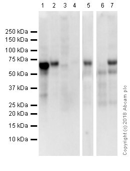
All lanes : Anti-Estrogen Receptor alpha antibody [E115] - ChIP Grade (ab32063) at 1/1000 dilution
Lane 1 : MCF7 (Human breast adenocarcinoma epithelial cell). Whole cell lysates
Lane 2 : T-47D (human mammary gland ductal carcinoma epithelial cell). Whole cell lysates
Lane 3 : MDA-MB231 (Human breast adenocarcinoma epithelial cell) Whole cell lysates (Negative control)
Lane 4 : HepG2 (Human hepatocellular carcinoma epithelial cell) Whole cell lysates (Negative control)
Lane 5 : Human uterus whole tissue lysate
Lane 6 : Human ovary whole tissue lysate
Lane 7 : Human ovary cancer whole tissue lysate
Lysates/proteins at 20 µg per lane.
Secondary
All lanes : Goat Anti-Rabbit IgG H&L (HRP) (ab97051) at 1/20000 dilution
Exposure time: 50 secondsBlocking and diluting buffer: 5% NFDM/TBST.
-
 Immunohistochemistry (Formalin/PFA-fixed paraffin-embedded sections) - Goat Anti-Rabbit IgG H&L (HRP) (ab97051)Immunohistochemical staining of paraffin embedded human endometrial carcinoma with purified ab32063 at a working dilution of 1 in 200. The secondary antibody used is ab97051, a HRP goat anti-rabbit IgG (H+L), at 1/500. The sample is counter-stained with hematoxylin. Antigen retrieval was perfomed using Tris-EDTA buffer, pH 9.0.
 -
 Western blot - Goat Anti-Rabbit IgG H&L (HRP) (ab97051) Mamedov et al PLoS One. 2017 Aug 21;12(8):e0183589. doi: 10.1371/journal.pone.0183589. eCollection 2017. Fig 4. Reproduced under the Creative Commons license http://creativecommons.org/licenses/by/4.0/
Western blot analysis of co-expression Bacillus anthracis PA83 (A), Pfs48/45 (B) and Pfs48/45-10C with bacterial Endo H or PNGase F in N. benthamiana plants.
(A) Western blot analysis of co-expression of PA83. Lanes: 1- N. benthamiana plant was infiltrated with pBI-PA83 construct, for the production of glycosylated PA83, 2,3- N. benthamiana plants were infiltrated with combinations of the pBI-Endo H/pBI-PA83 or pBI-PNGase F/pBI-PA83 constructs, for the production of Endo H (2) or PNGase F (3) deglycosylated PA83 proteins.
(B) Western blot analysis of co-expression of Pfs48/45. Lanes: 1-N. benthamiana plant was infiltrated with pEAQ-Pfs48/45 construct for the production of glycosylated Pfs48/45;2,3- N. benthamiana plants were infiltrated with combinations of the pBI-Endo H/pEAQ-Pfs48/45 or pBI-PNGase F/pEAQ-Pfs48/45constructs for the production of Endo H (2) and PNGase F (3) deglycosylated Pfs48/45 proteins.
(C) Western blot analysis of co-expression of Pfs48/45-10C. Lanes: 1- N. benthamiana plant was infiltrated with pEAQ-Pfs48/45-10C construct for the production of glycosylated Pfs48/45-10C; 2,3- N. benthamiana plants were infiltrated with combinations of the pBI-Endo H/pEAQ-Pfs48/45 or pBI-PNGase F/pEAQ-Pfs48/45constructs for the production of Endo H (2) and PNGase F (3) deglycosylated Pfs48/45-10C proteins. gPA83- glycosylated PA83; dPA83- deglycosylated PA83; gPfs48/45: glycosylated Pfs48/45; dPfs48/45: deglycosylated Pfs48/45; gPfs48/45-10C: glycosylated Pfs48/45-10C; dPfs48/45-10C: deglycosylated Pfs48/45-10C.
M: MagicMark XP Western Protein Standard. PA83 proteins were detected using the anti-Bacillus anthracis protective antigen antibody BAP0101 (Cat. No. ab1988, Abcam); Ps48/45, Endo H or PNGase F proteins were detected using the anti-FLAG antibody. Pfs48/45-10C protein was detected using the purified anti-His Tag antibody.
-
 Immunohistochemistry (Formalin/PFA-fixed paraffin-embedded sections) - Goat Anti-Rabbit IgG H&L (HRP) (ab97051)
IHC image of beta Actin staining in normal human colon, formalin-fixed and paraffin-embedded tissue*.
The section was pre-treated using pressure cooker heat mediated antigen retrieval with sodium citrate buffer (pH 6) for 30mins. The section was incubated with ab8227, 3 µg/ml overnight at +4°C. An HRP-conjugated secondary (ab97051, 1/2000 dilution) was used for 1hr at room temperature. The section was counterstained with hematoxylin and mounted with DPX.
The inset negative control image is secondary-only at 1/500 dilution.
*Tissue obtained from the Human Research Tissue Bank, supported by the NIHR Cambridge Biomedical Research Centre
-
 Western blot - Goat Anti-Rabbit IgG H&L (HRP) (ab97051) This image is courtesy of an anonymous Abreview.Anti-LAMP2 antibody (ab37024) at 1/1000 dilution + Mouse brain whole tissue lysate at 30 µg
Secondary
Goat Anti-Rabbit IgG H&L (HRP) (ab97051) at 1/5000 dilution
Developed using the ECL technique.
Performed under reducing conditions.
Exposure time: 1 minute10 % gel. Blocked with 5% BSA for 2 hours at 25°C.
Incubated with the primary antibody for 1 hour in TBS-tween at 25°C.
-
All lanes : Anti-beta Actin antibody (ab8227) at 1 µg/ml
All lanes : HeLa (Human epithelial carcinoma cell line) Whole Cell Lysate
Lysates/proteins at 10 µg per lane.
Secondary
Lanes 1-2 : Rabbit polyclonal to GNAT2 (ab97501) at 1/2000 dilution
Lanes 3-4 : Goat Anti-Rabbit IgG H&L (HRP) (ab97051) at 1/10000 dilution
Lanes 5-6 : Goat Anti-Rabbit IgG H&L (HRP) (ab97051) at 1/20000 dilution
Developed using the ECL technique.
Performed under reducing conditions.
Exposure time: 10 seconds


