ATP Assay Kit (Colorimetric/Fluorometric) (ab83355)
 (ab83355)")
Key features and details
- Assay type: Quantitative
- Detection method: Colorimetric/Fluorometric
- Platform: Microplate reader
- Assay time: 1 hr
- Sample type: Cell Lysate, Other biological fluids, Plasma, Serum, Tissue Extracts, Urine
- Sensitivity: 1 µM
Overview
-
Product name
ATP Assay Kit (Colorimetric/Fluorometric)
See all ATP kits -
Detection method
Colorimetric/Fluorometric -
Sample type
Urine, Serum, Plasma, Other biological fluids, Tissue Extracts, Cell Lysate -
Assay type
Quantitative -
Sensitivity
Assay time
1h 00mProduct overview
ATP Assay Kit (Colorimetric/Fluorometric) (ab83355) use a robust, simple method; the ATP assay protocol relies on the phosphorylation of glycerol to generate a product that is easily quantified by colorimetric (ODmax = 570 nm) or fluorometric (Ex/Em = 535/587 nm) methods.
This kit can detect as low as 1 µM of ATP in various samples.
ATP assay protocol summary:
- add samples (deproteinized) and standards to wells
- add reaction mix and incubate for 30 min at room temp
- analyze with microplate readerChinese protocol available. See protocols section below.
If you require a more sensitive product, we recommend Luminescent ATP Detection Assay Kit (ab113849), which can detect as low as 1 pM of ATP.
Notes
Related assays
Review the cell health assay guide to learn about kits to perform a cell viability assay, cytotoxicity assay and cell proliferation assay.
Review the metabolism assay guide to learn about assays for metabolites, metabolic enzymes, mitochondrial function, and oxidative stress, and also about how to assay metabolic function in live cells using your plate reader.
How other researchers have used ATP Assay Kit ab83355
This ATP assay kit has been used in publications in a variety of sample types, including:
- Human: cell culture lysates1, primary monocyte cell culture lysates2, HCT116 cell culture supernatants3
- Mouse: heart tissue4, liver5, C2C12 and L929 cell lysates6, primary thymocyte cell culture lysates7, cardiac tissue8
- Rat: primary hippocampal neuron cell culture lysates9, liver tissue10, skeletal muscle11
- Pig: kidney cell culture lysates12, heart tissue13
- C elegans tissue14
- Chlamydomonas reinhardtii algae15References: 1 - Civallero M et al 2017, Na JY et al 2018, 2Gkirtzimanaki K et al 2018, 3Yang et al 2018; 4 - Singh SP et al 2018; 5 - Han SJ et al 2018; 6 - Alhindi Y et al 2019, Gregorczyk KP et al 2018; 7 - Simula L et al 2018; 8 - Litt MJ et al 2017, Samokhvalov V et al 2018; 9 - Zhao X et al 2018; 10 - Jing R et al 2018; 11 - Trinchese G et al 2018; 12 - Zou X et al 2018; 13 - Yuan F et al 2018; 14 - Pandey et al 2018; 15 - Ramanan R et al 2018
Platform
Microplate readerProperties
-
Storage instructions
Store at -20°C. Please refer to protocols. -
Components Identifier 100 tests ATP Assay Buffer WM 1 x 25ml ATP Converter (lyophilized) Blue 1 vial ATP Probe (in DMSO) Red 1 x 200µl ATP Standard (1 µmol; lyophilized) Yellow 1 vial Developer Mix (lyophilized) Green 1 vial -
Research areas
-
Alternative names
- Adenosine 5' triphosphate
Properties
-
Storage instructions
Store at -20°C. Please refer to protocols. -
Components Identifier 100 tests ATP Assay Buffer WM 1 x 25ml ATP Converter (lyophilized) Blue 1 vial ATP Probe (in DMSO) Red 1 x 200µl ATP Standard (1 µmol; lyophilized) Yellow 1 vial Developer Mix (lyophilized) Green 1 vial -
Research areas
-
Alternative names
- Adenosine 5' triphosphate
Images
-
ATP levels in Pancreatic Islet Image courtesy of Mrs. Fotini Mouth
Ab83355 was used to determin ATP levels in rat pancreas islets as an ischemic marker to predict transplantation outcomes. We extracted ATP from fresh pancreas that have undergo different time of cold ischemia : 0, 2, 4, 6, 8 and 10h and in situ. ATP were extracted in Perchloric acid (PCA-2M) and grind using a Polytron. PCA were removed using potassium hydroxide (KOH – 2M) and pH was adjust around 7-8. Samples were conserved at -80°C before utilization.
-
 ATP assay used to study mitochondrial disfunction after C. rodentium infection in mice Image courtesy of Maiti A K et al. PLoS One. 2018; 13(9): e0204567. doi: 10.1371/journal.pone.0204567
Maiti AK et al (2018) used ATP assay kit ab83355 to measure mitochondrial ATP generation in an in vitro mouse intestinal model treated with cytokines in the presence and absence of VIP (vasoactive intestinal peptide). VIP was induced by C. rodentium infection and cytokines.
-
 ATP assay performed with ab83355 Sanokawa-Akakura R et al., PLoS One 9(9), fig4b. doi: 10.1371/journal.pone.0108537 Reproduced under the Creative Commons license http://creativecommons.org/licenses/by/4.0/
The chart shows a comparison of ATP levels of HepG2 treated with 0, 10 and 100 µM for 48 hours, DRH2O2 W1 (damage recovered cells using hydrogen peroxide with a recovery time of one week) HepG2 cells and MDA-MB-231 cells treated with 0, 10 and 100 µM of NaHS for 48 hours. Data is shown as percent of ATP levels in untreated cells. ATP levels were determined using ATP assay kit (ab83355).
-
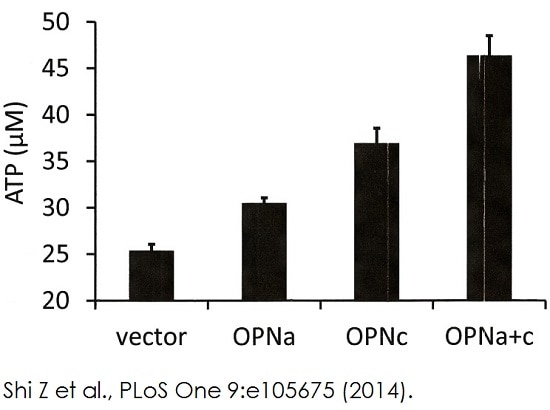 ATP assay performed with ab83355 Image from Shi Z et al., PLoS One 9(8), Fig 2 doi: 10.1371/journal.pone.0105675. Reproduced under the Creative Commons license http://creativecommons.org/licenses/by/4.0/
MCF-7 cells are transfected with vector, osteopontin-a, osteopontin-c or osteopontin -a plus -c. Cells are plated on poly-HEMA and seeded at 4 x 10⁵ cells per well and incubated for two days under standard culture conditions. ATP levels are measured using ATP assay kit (ab83355).
-
 ATP assay performed with ab83355Example of fluorometric ATP assay standard curve.
 -
 ATP levels measured in mouse colon tissue Image courtesy of Maiti A K et al. Sci Rep. 2015; 5: 1543. doi: 10.1038/srep15434. Reproduced under the Creative Commons License http://creativecommons.org/licenses/by/4.0/
Maiti AK et al. (2015) used ATP assay kit ab113852 to measure mitochondrial ATP generation in murine distal colon after C. rodentium infection.
-
 ATP assay performed with ab83355Example of colorimetric ATP assay standard curve.
 -
 ATP assay performed with ab83355
Quantitation of ATP in fish liver (2.5µl of 10 times diluted sample), fish muscle (5µl of 10 times diluted sample) and Jurkat cell lysate (5 ul) using fluorometric assay. Samples were spiked with known amounts of ATP (300pmol).


