Anti-USP2 antibody (ab66556)
")
Key features and details
- Rabbit polyclonal to USP2
- Suitable for: WB, ICC/IF
- Reacts with: Human
- Isotype: IgG
Overview
-
Product name
Anti-USP2 antibody
See all USP2 primary antibodies -
Description
Rabbit polyclonal to USP2 -
Host species
Rabbit -
Tested Applications & Species
See all applications and species dataApplication Species ICC/IF HumanWB Human
-
Immunogen
Synthetic peptide corresponding to Human USP2 aa 200-300 conjugated to keyhole limpet haemocyanin.
(Peptide available asab66555) -
Positive control
- This antibody gave a positive signal in the following Human Tissue Lysates: Pancreas, Prostate, Prostate Tumor
Properties
-
Form
Liquid -
Storage instructions
Shipped at 4°C. Store at +4°C short term (1-2 weeks). Upon delivery aliquot. Store at -20°C or -80°C. Avoid freeze / thaw cycle. -
Storage buffer
pH: 7.40
Preservative: 0.02% Sodium azide
Constituent: PBS
Batches of this product that have a concentration Concentration information loading...
Purity
Immunogen affinity purifiedClonality
PolyclonalIsotype
IgGResearch areas
Associated products
-
Compatible Secondaries
-
Isotype control
-
Recombinant Protein
Applications
The Abpromise guarantee
Our Abpromise guarantee covers the use of ab66556 in the following tested applications.
The application notes include recommended starting dilutions; optimal dilutions/concentrations should be determined by the end user.
GuaranteedTested applications are guaranteed to work and covered by our Abpromise guarantee.
PredictedPredicted to work for this combination of applications and species but not guaranteed.
IncompatibleDoes not work for this combination of applications and species.
Application Species ICC/IF HumanWB HumanAll applications RatChimpanzeeRhesus monkeyApplication Abreviews Notes WB Use a concentration of 1 µg/ml. Detects a band of approximately 68 kDa (predicted molecular weight: 68 kDa).ICC/IF Use a concentration of 5 µg/ml.Notes WB
Use a concentration of 1 µg/ml. Detects a band of approximately 68 kDa (predicted molecular weight: 68 kDa).ICC/IF
Use a concentration of 5 µg/ml.Target
-
Function
Hydrolase that deubiquitinates polyubiquitinated target proteins such as MDM2, MDM4 and CCND1. Isoform 1 and isoform 4 possess both ubiquitin-specific peptidase and isopeptidase activities. Deubiquitinates MDM2 without reversing MDM2-mediated p53/TP53 ubiquitination and thus indirectly promotes p53/TP53 degradation and limits p53 activity. Has no deubiquitinase activity against p53/TP53. Prevents MDM2-mediated degradation of MDM4. Plays a role in the G1/S cell-cycle progression in normal and cancer cells. Plays a role in the regulation of myogenic differentiation of embryonic muscle cells. -
Tissue specificity
Expressed in mesangial cells of the kidney and in different types of glomerulonephritides (at protein level). -
Sequence similarities
Belongs to the peptidase C19 family. USP2 subfamily. -
Domain
The different N-terminus extensions of isoform 1 and isoform 4 determine their respective subcellular localization and differentiel effect on myoblast fusion and accumulation of muscle-specific proteins. The different N-terminus extensions of isoform 1 and isoform 4 are not essential for their catalytic activity. -
Cellular localization
Nucleus and Cytoplasm. Cytoplasm > perinuclear region. Localizes in the spermatid head in late-elongating spermatids in the thin area between the outer acrosomal membrane and the plasma membrane. - Information by UniProt
-
Database links
- Entrez Gene: 9099 Human
- Entrez Gene: 115771 Rat
- Omim: 604725 Human
- SwissProt: O75604 Human
- SwissProt: Q5U349 Rat
- Unigene: 524085 Human
- Unigene: 203590 Rat
-
Alternative names
- 41 kDa ubiquitin specific protease antibody
- 41 kDa ubiquitin-specific protease antibody
- Deubiquitinating enzyme 2 antibody
see all
Images
-
Western blot - Anti-USP2 antibody (ab66556)All lanes : Anti-USP2 antibody (ab66556) at 1 µg/ml
Lane 1 : Human pancreas tissue lysate - total protein (ab29816)
Lane 2 : Human prostate tissue lysate - total protein (ab30304)
Lane 3 : Prostate Tumor Lysate Tissue Lysate
Lysates/proteins at 10 µg per lane.
Secondary
All lanes : Goat polyclonal to Rabbit IgG - H&L - Pre-Adsorbed (HRP) at 1/3000 dilution
Developed using the ECL technique.
Performed under reducing conditions.
Predicted band size: 68 kDa
Observed band size: 68 kDa
Additional bands at: 30 kDa, 56 kDa. We are unsure as to the identity of these extra bands. -
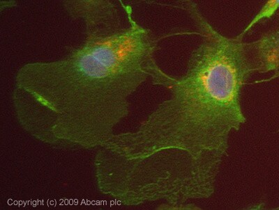 Immunocytochemistry/ Immunofluorescence - Anti-USP2 antibody (ab66556)ICC/IF image of ab66556 stained HepG2 cells. The cells were 4% PFA fixed (10 min) and then incubated in 1%BSA / 10% normal Goat serum / 0.3M glycine in 0.1% PBS-Tween for 1h to permeabilise the cells and block non-specific protein-protein interactions. The cells were then incubated with the antibody (ab66556, 5µg/ml) overnight at +4°C. The secondary antibody (green) was Alexa Fluor® 488 Goat anti-Rabbit IgG (H+L) used at a 1/1000 dilution for 1h. Alexa Fluor® 594 WGA was used to label plasma membranes (red) at a 1/200 dilution for 1h. DAPI was used to stain the cell nuclei (blue) at a concentration of 1.43µM. This antibody also gave a positive result in 100% Methanol fixed (5 min) HepG2 cells at 5µg/ml.
Protocols
Datasheets and documents
References (1)
ab66556 has been referenced in 1 publication.
- Zhou Y et al. Hepatocellular carcinoma-derived exosomal miRNA-21 contributes to tumor progression by converting hepatocyte stellate cells to cancer-associated fibroblasts. J Exp Clin Cancer Res 37:324 (2018). PubMed: 30591064
Images
-
Western blot - Anti-USP2 antibody (ab66556)All lanes : Anti-USP2 antibody (ab66556) at 1 µg/ml
Lane 1 : Human pancreas tissue lysate - total protein (ab29816)
Lane 2 : Human prostate tissue lysate - total protein (ab30304)
Lane 3 : Prostate Tumor Lysate Tissue Lysate
Lysates/proteins at 10 µg per lane.
Secondary
All lanes : Goat polyclonal to Rabbit IgG - H&L - Pre-Adsorbed (HRP) at 1/3000 dilution
Developed using the ECL technique.
Performed under reducing conditions.
Predicted band size: 68 kDa
Observed band size: 68 kDa
Additional bands at: 30 kDa, 56 kDa. We are unsure as to the identity of these extra bands.
-
Immunocytochemistry/ Immunofluorescence - Anti-USP2 antibody (ab66556)ICC/IF image of ab66556 stained HepG2 cells. The cells were 4% PFA fixed (10 min) and then incubated in 1%BSA / 10% normal Goat serum / 0.3M glycine in 0.1% PBS-Tween for 1h to permeabilise the cells and block non-specific protein-protein interactions. The cells were then incubated with the antibody (ab66556, 5µg/ml) overnight at +4°C. The secondary antibody (green) was Alexa Fluor® 488 Goat anti-Rabbit IgG (H+L) used at a 1/1000 dilution for 1h. Alexa Fluor® 594 WGA was used to label plasma membranes (red) at a 1/200 dilution for 1h. DAPI was used to stain the cell nuclei (blue) at a concentration of 1.43µM. This antibody also gave a positive result in 100% Methanol fixed (5 min) HepG2 cells at 5µg/ml.

