Anti-Talin 1 antibody (ab71333)
Key features and details
- Rabbit polyclonal to Talin 1
- Suitable for: ICC/IF, IHC-P, WB
- Reacts with: Mouse, Human
- Isotype: IgG
Overview
-
Product name
Anti-Talin 1 antibody
See all Talin 1 primary antibodies -
Description
Rabbit polyclonal to Talin 1 -
Host species
Rabbit -
Tested Applications & Species
See all applications and species dataApplication Species ICC/IF MouseHumanIHC-P HumanWB Human -
Immunogen
Synthetic peptide corresponding to Human Talin 1 aa 1650-1750 (internal sequence) conjugated to keyhole limpet haemocyanin.
(Peptide available asab71332) -
General notes
The Life Science industry has been in the grips of a reproducibility crisis for a number of years. Abcam is leading the way in addressing the problem with our range of recombinant monoclonal antibodies and knockout edited cell lines for gold-standard validation.
One factor contributing to the crisis is the use of antibodies that are not suitable. This can lead to misleading results and the use of incorrect data informing project assumptions and direction. To help address this challenge, we have introduced an application and species grid on our primary antibody datasheets to make it easy to simplify identification of the right antibody for your needs.
Learn more here.
Properties
-
Form
Liquid -
Storage instructions
Shipped at 4°C. Store at +4°C short term (1-2 weeks). Upon delivery aliquot. Store at -20°C or -80°C. Avoid freeze / thaw cycle. -
Storage buffer
pH: 7.40
Preservative: 0.02% Sodium azide
Constituent: PBS
Batches of this product that have a concentration Concentration information loading...
Purity
Immunogen affinity purifiedClonality
PolyclonalIsotype
IgGResearch areas
Associated products
-
Compatible Secondaries
-
Isotype control
Applications
The Abpromise guarantee
Our Abpromise guarantee covers the use of ab71333 in the following tested applications.
The application notes include recommended starting dilutions; optimal dilutions/concentrations should be determined by the end user.
GuaranteedTested applications are guaranteed to work and covered by our Abpromise guarantee.
PredictedPredicted to work for this combination of applications and species but not guaranteed.
IncompatibleDoes not work for this combination of applications and species.
Application Species ICC/IF MouseHumanIHC-P HumanWB HumanAll applications RatChickenApplication Abreviews Notes ICC/IF Use a concentration of 1 µg/ml.IHC-P (1) Use a concentration of 5 µg/ml.WB (2) Use a concentration of 1 µg/ml. Detects a band of approximately 270 kDa (predicted molecular weight: 270 kDa).Notes ICC/IF
Use a concentration of 1 µg/ml.IHC-P
Use a concentration of 5 µg/ml.WB
Use a concentration of 1 µg/ml. Detects a band of approximately 270 kDa (predicted molecular weight: 270 kDa).Target
-
Function
Probably involved in connections of major cytoskeletal structures to the plasma membrane. High molecular weight cytoskeletal protein concentrated at regions of cell-substratum contact and, in lymphocytes, at cell-cell contacts. -
Sequence similarities
Contains 1 FERM domain.
Contains 1 I/LWEQ domain. -
Cellular localization
Cell projection, ruffle membrane. Cytoplasm, cytoskeleton. Cell surface. Cell junction, focal adhesion. Colocalizes with LAYN at the membrane ruffles. Localized preferentially in focal adhesions than fibrillar adhesions (By similarity). - Information by UniProt
-
Database links
- Entrez Gene: 395194 Chicken
- Entrez Gene: 7094 Human
- Entrez Gene: 21894 Mouse
- Entrez Gene: 313494 Rat
- Omim: 186745 Human
- SwissProt: P54939 Chicken
- SwissProt: Q9Y490 Human
- SwissProt: P26039 Mouse
see all -
Alternative names
- ILWEQ antibody
- Talin 1 antibody
- Talin antibody
see all
Images
-
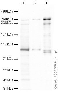
All lanes : Anti-Talin 1 antibody (ab71333) at 1 µg/ml
Lane 1 : Jurkat (Human T cell lymphoblast-like cell line) Whole Cell Lysate
Lane 2 : Ramos (Human Burkitt's lymphoma cell line) Whole Cell Lysate
Lane 3 : MEF1 (Mouse embryonic fibroblast cell line) Whole Cell Lysate
Lysates/proteins at 10 µg per lane.
Secondary
All lanes : Goat polyclonal to Rabbit IgG - H&L - Pre-Adsorbed (HRP) at 1/3000 dilution
Performed under reducing conditions.
Predicted band size: 270 kDa
Observed band size: 270 kDa
Additional bands at: 100 kDa, 230 kDa. We are unsure as to the identity of these extra bands.
Exposure time: 8 minutes -
 Immunohistochemistry (Formalin/PFA-fixed paraffin-embedded sections) - Anti-Talin 1 antibody (ab71333)IHC image of Talin 1 staining in Human Normal Kidney FFPE section, performed on a BondTM system using the standard protocol F. The section was pre-treated using heat mediated antigen retrieval with sodium citrate buffer (pH6, epitope retrieval solution 1) for 20 mins. The section was then incubated with ab71333, 5µg/ml, for 15 mins at room temperature and detected using an HRP conjugated compact polymer system. DAB was used as the chromogen. The section was then counterstained with haematoxylin and mounted with DPX.
 -
 Immunocytochemistry/ Immunofluorescence - Anti-Talin 1 antibody (ab71333)Courtesy of Dr. Shaohua Li, UMDNJ-Robert Wood Johnson Medical School
Image: Courtesy of Dr. Shaohua Li, UMDNJ-Robert Wood Johnson Medical School
Sample: mouse endoderm cell
Preparation:
Fix in 3% PFA in PBS for 30 min at RT
Primary antibody: Rabbit anti-talin (ab71333), 1:100
Secondary antibody: Goat polyclonal Secondary Antibody to Rabbit IgG - H&L (Cy3® 488) preadsorbed (ab97075), 1:100
Nuclear counterstain: DAPI
Rhodamine-phalloidin, 1:100
Nuclei were counterstained with DAPI
-
ab71333 stained in HeLa cells. Cells were fixed with 4% paraformaldehyde (10 min) at room temperature and incubated with PBS containing 10% goat serum, 0.3 M glycine, 1% BSA and 0.1% Triton for 1h at room temperature to permeabilise the cells and block non-specific protein-protein interactions. The cells were then incubated with the antibody ab71333 at 5µg/ml and ab7291 (Mouse monoclonal to alpha Tubulin - Loading Control) used at a 1/1000 dilution overnight at +4°C. The secondary antibodies were ab150081, Goat Anti-Rabbit IgG H&L (Alexa Fluor® 488) preadsorbed, (pseudo-colored green) and ab150120, Goat polyclonal Secondary Antibody to Mouse IgG - H&L (Alexa Fluor® 594) preadsorbed, (colored red), both used at a 1/1000 dilution for 1 hour at room temperature. DAPI was used to stain the cell nuclei (colored blue) at a concentration of 1.43 µM for 1hour at room temperature.
-
All lanes : Anti-Talin 1 antibody (ab71333) at 1/1000 dilution
Lane 1 : Human platelets
Lane 2 : Mouse platelets
Lysates/proteins at 20 µg per lane.
Secondary
All lanes : Goat polyclonal to rabbit IgG conjugated to IRDye 800CW (undiluted)
Performed under reducing conditions.
Predicted band size: 270 kDa
Exposure time: 1 minute
Detection method: Licor System
Protocols
Datasheets and documents
-
SDS download
-
Datasheet download
References (14)
ab71333 has been referenced in 14 publications.
- Gao J et al. Long noncoding RNA LINC00488 functions as a ceRNA to regulate hepatocellular carcinoma cell growth and angiogenesis through miR-330-5. Dig Liver Dis N/A:N/A (2019). PubMed: 31005556
- Ji L et al. Talin1 knockdown prohibits the proliferation and migration of colorectal cancer cells via the EMT signaling pathway. Oncol Lett 18:5408-5416 (2019). PubMed: 31612049
- Ghosh M et al. CD13 tethers the IQGAP1-ARF6-EFA6 complex to the plasma membrane to promote ARF6 activation, ß1 integrin recycling, and cell migration. Sci Signal 12:N/A (2019). PubMed: 31040262
- Liang Y et al. Downregulation of Dock1 and Elmo1 suppresses the migration and invasion of triple-negative breast cancer epithelial cells through the RhoA/Rac1 pathway. Oncol Lett 16:3481-3488 (2018). PubMed: 30127952
- Tanner MR et al. KCa1.1 channels regulate ß1 integrin function and cell adhesion in rheumatoid arthritis fibroblast-like synoviocytes. FASEB J N/A:N/A (2017). PubMed: 28428266
Images
-
All lanes : Anti-Talin 1 antibody (ab71333) at 1 µg/ml
Lane 1 : Jurkat (Human T cell lymphoblast-like cell line) Whole Cell Lysate
Lane 2 : Ramos (Human Burkitt's lymphoma cell line) Whole Cell Lysate
Lane 3 : MEF1 (Mouse embryonic fibroblast cell line) Whole Cell Lysate
Lysates/proteins at 10 µg per lane.
Secondary
All lanes : Goat polyclonal to Rabbit IgG - H&L - Pre-Adsorbed (HRP) at 1/3000 dilution
Performed under reducing conditions.
Predicted band size: 270 kDa
Observed band size: 270 kDa
Additional bands at: 100 kDa, 230 kDa. We are unsure as to the identity of these extra bands.
Exposure time: 8 minutes
-
 Immunohistochemistry (Formalin/PFA-fixed paraffin-embedded sections) - Anti-Talin 1 antibody (ab71333)IHC image of Talin 1 staining in Human Normal Kidney FFPE section, performed on a BondTM system using the standard protocol F. The section was pre-treated using heat mediated antigen retrieval with sodium citrate buffer (pH6, epitope retrieval solution 1) for 20 mins. The section was then incubated with ab71333, 5µg/ml, for 15 mins at room temperature and detected using an HRP conjugated compact polymer system. DAB was used as the chromogen. The section was then counterstained with haematoxylin and mounted with DPX.
 -
 Immunocytochemistry/ Immunofluorescence - Anti-Talin 1 antibody (ab71333) Courtesy of Dr. Shaohua Li, UMDNJ-Robert Wood Johnson Medical School
Image: Courtesy of Dr. Shaohua Li, UMDNJ-Robert Wood Johnson Medical School
Sample: mouse endoderm cell
Preparation:
Fix in 3% PFA in PBS for 30 min at RT
Primary antibody: Rabbit anti-talin (ab71333), 1:100
Secondary antibody: Goat polyclonal Secondary Antibody to Rabbit IgG - H&L (Cy3® 488) preadsorbed (ab97075), 1:100
Nuclear counterstain: DAPI
Rhodamine-phalloidin, 1:100
Nuclei were counterstained with DAPI
-
ab71333 stained in HeLa cells. Cells were fixed with 4% paraformaldehyde (10 min) at room temperature and incubated with PBS containing 10% goat serum, 0.3 M glycine, 1% BSA and 0.1% Triton for 1h at room temperature to permeabilise the cells and block non-specific protein-protein interactions. The cells were then incubated with the antibody ab71333 at 5µg/ml and ab7291 (Mouse monoclonal to alpha Tubulin - Loading Control) used at a 1/1000 dilution overnight at +4°C. The secondary antibodies were ab150081, Goat Anti-Rabbit IgG H&L (Alexa Fluor® 488) preadsorbed, (pseudo-colored green) and ab150120, Goat polyclonal Secondary Antibody to Mouse IgG - H&L (Alexa Fluor® 594) preadsorbed, (colored red), both used at a 1/1000 dilution for 1 hour at room temperature. DAPI was used to stain the cell nuclei (colored blue) at a concentration of 1.43 µM for 1hour at room temperature.
-
All lanes : Anti-Talin 1 antibody (ab71333) at 1/1000 dilution
Lane 1 : Human platelets
Lane 2 : Mouse platelets
Lysates/proteins at 20 µg per lane.
Secondary
All lanes : Goat polyclonal to rabbit IgG conjugated to IRDye 800CW (undiluted)
Performed under reducing conditions.
Predicted band size: 270 kDa
Exposure time: 1 minute
Detection method: Licor System


