Anti-STEAP3 antibody (ab104654)
Key features and details
- Rabbit polyclonal to STEAP3
- Suitable for: WB, ICC/IF
- Reacts with: Mouse, Human
- Isotype: IgG
Overview
-
Product name
Anti-STEAP3 antibody
See all STEAP3 primary antibodies -
Description
Rabbit polyclonal to STEAP3 -
Host species
Rabbit -
Tested Applications & Species
See all applications and species dataApplication Species ICC/IF HumanWB Mouse -
Immunogen
Synthetic peptide corresponding to Mouse STEAP3 aa 150-250 conjugated to keyhole limpet haemocyanin.
(Peptide available asab130906) -
Positive control
- This antibody gave a positive signal in the following tissue lysates: Mouse Liver; Mouse Brain; Mouse Heart. This antibody gave a positive result in IF in the following formaldehyde fixed cell lines: JEG3.
Properties
-
Form
Liquid -
Storage instructions
Shipped at 4°C. Store at +4°C short term (1-2 weeks). Upon delivery aliquot. Store at -20°C or -80°C. Avoid freeze / thaw cycle. -
Storage buffer
pH: 7.40
Preservative: 0.02% Sodium azide
Constituent: PBS
Batches of this product that have a concentration Concentration information loading...
Purity
Immunogen affinity purifiedClonality
PolyclonalIsotype
IgGResearch areas
Associated products
-
Compatible Secondaries
-
Isotype control
Applications
The Abpromise guarantee
Our Abpromise guarantee covers the use of ab104654 in the following tested applications.
The application notes include recommended starting dilutions; optimal dilutions/concentrations should be determined by the end user.
GuaranteedTested applications are guaranteed to work and covered by our Abpromise guarantee.
PredictedPredicted to work for this combination of applications and species but not guaranteed.
IncompatibleDoes not work for this combination of applications and species.
Application Species ICC/IF HumanWB MouseApplication Abreviews Notes WB Use a concentration of 1 µg/ml. Detects a band of approximately 55 kDa (predicted molecular weight: 55 kDa).ICC/IF Use a concentration of 1 µg/ml.Notes WB
Use a concentration of 1 µg/ml. Detects a band of approximately 55 kDa (predicted molecular weight: 55 kDa).ICC/IF
Use a concentration of 1 µg/ml.Target
-
Function
Endosomal ferrireductase required for efficient transferrin-dependent iron uptake in erythroid cells. Participates in erythroid iron homeostasis by reducing Fe(3+) to Fe(2+). Can also reduce of Cu(2+) to Cu(1+), suggesting that it participates in copper homeostasis. Uses NADP(+) as acceptor. May play a role downstream of p53/TP53 to interface apoptosis and cell cycle progression. Indirectly involved in exosome secretion by facilitating the secretion of proteins such as TCTP. -
Tissue specificity
Expressed in adult bone marrow, placenta, liver, skeletal muscle and pancreas. Down-regulated in hepatocellular carcinoma. -
Involvement in disease
Anemia, hypochromic microcytic, with iron overload 2 -
Sequence similarities
Belongs to the STEAP family.
Contains 1 ferric oxidoreductase domain. -
Post-translational
modificationsProteolytically cleaved by RHBDL4/RHBDD1. RHBDL4/RHBDD1-induced cleavage occurs at multiple sites in a glycosylation-independent manner.
Glycosylated. -
Cellular localization
Endosome membrane. Localizes to vesicular-like structures at the plasma membrane and around the nucleus. - Information by UniProt
-
Database links
- Entrez Gene: 55240 Human
- Entrez Gene: 68428 Mouse
- Omim: 609671 Human
- SwissProt: Q658P3 Human
- SwissProt: Q8CI59 Mouse
- Unigene: 647822 Human
- Unigene: 181033 Mouse
-
Alternative names
- 1010001D01Rik antibody
- Dudlin 2 antibody
- Dudlin2 antibody
see all
Images
-
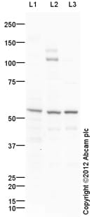
All lanes : Anti-STEAP3 antibody (ab104654) at 1 µg/ml
Lane 1 : Liver (Mouse) Tissue Lysate
Lane 2 : Brain (Mouse) Tissue Lysate
Lane 3 : Heart (Mouse) Tissue Lysate
Lysates/proteins at 10 µg per lane.
Secondary
All lanes : Goat Anti-Rabbit IgG H&L (HRP) preadsorbed (ab97080) at 1/5000 dilution
Developed using the ECL technique.
Performed under reducing conditions.
Predicted band size: 55 kDa
Observed band size: 55 kDa
Additional bands at: 105 kDa, 130 kDa, 46 kDa. We are unsure as to the identity of these extra bands.
Exposure time: 1 minute -
ICC/IF image of ab104654 stained JEG3 cells. The cells were 4% formaldehyde fixed (5 min) and then incubated in 1%BSA / 10% normal goat serum / 0.3M glycine in 0.1% PBS-Tween for 1h to permeabilise the cells and block non-specific protein-protein interactions. The cells were then incubated with the antibody ab104654 at 1µg/ml overnight at +4°C. The secondary antibody (green) was DyLight® 488 goat anti- rabbit (ab96899) IgG (H+L) used at a 1/1000 dilution for 1h. Alexa Fluor® 594 WGA was used to label plasma membranes (red) at a 1/200 dilution for 1h. DAPI was used to stain the cell nuclei (blue) at a concentration of 1.43µM.
Protocols
Datasheets and documents
References (0)
ab104654 has not yet been referenced specifically in any publications.
Images
-
All lanes : Anti-STEAP3 antibody (ab104654) at 1 µg/ml
Lane 1 : Liver (Mouse) Tissue Lysate
Lane 2 : Brain (Mouse) Tissue Lysate
Lane 3 : Heart (Mouse) Tissue Lysate
Lysates/proteins at 10 µg per lane.
Secondary
All lanes : Goat Anti-Rabbit IgG H&L (HRP) preadsorbed (ab97080) at 1/5000 dilution
Developed using the ECL technique.
Performed under reducing conditions.
Predicted band size: 55 kDa
Observed band size: 55 kDa
Additional bands at: 105 kDa, 130 kDa, 46 kDa. We are unsure as to the identity of these extra bands.
Exposure time: 1 minute
-
ICC/IF image of ab104654 stained JEG3 cells. The cells were 4% formaldehyde fixed (5 min) and then incubated in 1%BSA / 10% normal goat serum / 0.3M glycine in 0.1% PBS-Tween for 1h to permeabilise the cells and block non-specific protein-protein interactions. The cells were then incubated with the antibody ab104654 at 1µg/ml overnight at +4°C. The secondary antibody (green) was DyLight® 488 goat anti- rabbit (ab96899) IgG (H+L) used at a 1/1000 dilution for 1h. Alexa Fluor® 594 WGA was used to label plasma membranes (red) at a 1/200 dilution for 1h. DAPI was used to stain the cell nuclei (blue) at a concentration of 1.43µM.


