Anti-SREBP2 antibody (ab30682)
")
Key features and details
- Rabbit polyclonal to SREBP2
- Suitable for: ICC/IF, WB
- Reacts with: Rat, Human
- Isotype: IgG
Overview
-
Product name
Anti-SREBP2 antibody
See all SREBP2 primary antibodies -
Description
Rabbit polyclonal to SREBP2 -
Host species
Rabbit -
Tested applications
Suitable for: ICC/IF, WBmore details -
Species reactivity
Reacts with: Rat, Human
Predicted to work with: Guinea pig, Pig, Xenopus laevis, Chinese hamster
-
Immunogen
Synthetic peptide corresponding to Human SREBP2 aa 455-469.
Sequence:SPLLDDAKVKDEPDS
Database link: Q12772 -
Positive control
- ICC: HeLa cells. WB: Human fibroblast cell lysate, Rat testis supernatant, Human retina whole cell lysate.


Properties
-
Form
Liquid -
Storage instructions
Shipped at 4°C. Store at +4°C short term (1-2 weeks). Upon delivery aliquot. Store at -20°C. Avoid freeze / thaw cycle. -
Storage buffer
pH: 7.40
Preservative: 0.02% Sodium azide
Constituents: Tris buffered saline, 50% Glycerol, 0.1% BSA -
 Concentration information loading...
 -
Purity
Immunogen affinity purified -
Clonality
Polyclonal -
Isotype
IgG -
Research areas
- Epigenetics and Nuclear Signaling
- Transcription
- Domain Families
- HLH / Leucine Zipper
- HLH / Leucine Zipper
- Metabolism
- Pathways and Processes
- Metabolic signaling pathways
- Lipid and lipoprotein metabolism
- Lipid metabolism
Images
-
Western blot - Anti-SREBP2 antibody (ab30682) This image is courtesy of an Abreview by Dongil Kim.Anti-SREBP2 antibody (ab30682) at 1/1000 dilution + Human retina whole cell lysate at 30 µg
Secondary
HRP-conjugated goat anti-rabbit polyclonal at 1/1000 dilution
Developed using the ECL technique.
Performed under reducing conditions.
Predicted band size: 126 kDa
Additional bands at: 126, 55 kDa, 60 kDa (possible non-specific binding). We are unsure as to the identity of these extra bands.
Exposure time: 6 minutesBlocking: 7% milk for 60 minutes at 20oC.
-
 Immunocytochemistry/ Immunofluorescence - Anti-SREBP2 antibody (ab30682)
ICC/IF image of ab30682 stained HeLa cells. The cells were 4% PFA fixed (10 min) and then incubated in 1%BSA / 10% normal goat serum / 0.3M glycine in 0.1% PBS-Tween for 1h to permeabilise the cells and block non-specific protein-protein interactions. The cells were then incubated with the antibody (ab30682, 1µg/ml) overnight at +4°C. The secondary antibody (green) was Alexa Fluor® 488 goat anti-rabbit IgG (H+L) used at a 1/1000 dilution for 1h. Alexa Fluor® 594 WGA was used to label plasma membranes (red) at a 1/200 dilution for 1h. DAPI was used to stain the cell nuclei (blue) at a concentration of 1.43µM.
-
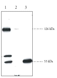 Western blot - Anti-SREBP2 antibody (ab30682)All lanes : Anti-SREBP2 antibody (ab30682) at 1/200 dilution
Lane 1 : Human fibroblast cell lysate
Lane 2 : Rat brown fat homogenate
Lane 3 : Rat testis supernatant
Lysates/proteins at 60 µg per lane.
Predicted band size: 126 kDa
Observed bands 126 kDa, 55kDa (cleaved form)


