Anti-SP1 antibody (ab227383)
")
Key features and details
- Rabbit polyclonal to SP1
- Suitable for: WB, IP, IHC-P, ChIP, ICC/IF
- Reacts with: Mouse, Human
- Isotype: IgG
Overview
-
Product name
Anti-SP1 antibody
See all SP1 primary antibodies -
Description
Rabbit polyclonal to SP1 -
Host species
Rabbit -
Tested Applications & Species
See all applications and species dataApplication Species ChIP HumanICC/IF HumanIHC-P MouseHumanIP HumanWB Human
-
Immunogen
Recombinant fragment within Human SP1 (internal sequence). The exact sequence is proprietary.
Database link: P08047 -
Positive control
- WB: HEK-293T and THP-1 whole cell lysate. ICC/IF: HeLa cells. IHC: HeLa and C2C12 xenografts. IP: THP-1 whole cell lysate. ChIP: HEK-293T chromatin extract.
-
General notes
The Life Science industry has been in the grips of a reproducibility crisis for a number of years. Abcam is leading the way in addressing the problem with our range of recombinant monoclonal antibodies and knockout edited cell lines for gold-standard validation.
One factor contributing to the crisis is the use of antibodies that are not suitable. This can lead to misleading results and the use of incorrect data informing project assumptions and direction. To help address this challenge, we have introduced an application and species grid on our primary antibody datasheets to make it easy to simplify identification of the right antibody for your needs.
Learn more here.
Properties
-
Form
Liquid -
Storage instructions
Shipped at 4°C. Store at +4°C short term (1-2 weeks). Upon delivery aliquot. Store at -20°C long term. Avoid freeze / thaw cycle. -
Storage buffer
pH: 7.00
Preservative: 0.025% Proclin 300
Constituents: 79% PBS, 20% Glycerol (glycerin, glycerine) -
 Concentration information loading...
 -
Purity
Immunogen affinity purified -
Clonality
Polyclonal -
Isotype
IgG -
Research areas
Images
-
Western blot - Anti-SP1 antibody (ab227383)All lanes : Anti-SP1 antibody (ab227383) at 1/10000 dilution
Lane 1 : Non-transfected HEK-293T (human epithelial cell line from embryonic kidney transformed with large T antigen) whole cell lysate
Lane 2 : SP1 shRNA transfected HEK-293T whole cell lysate
Lysates/proteins at 50 µg per lane.
Secondary
All lanes : HRP-conjugated anti-rabbit IgG
Developed using the ECL technique.
Performed under reducing conditions.
-
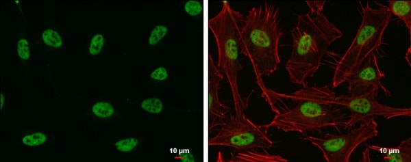 Immunocytochemistry/ Immunofluorescence - Anti-SP1 antibody (ab227383)
HeLa (human epithelial cell line from cervix adenocarcinoma) cells stained for SP1 (green) using ab227383 at 1/500 dilution in ICC/IF. Cells were fixed in 4% paraformaldehyde at RT for 15 minutes. Red: phalloidin, a cytoskeleton marker, diluted at 1/200.
-
 ChIP - Anti-SP1 antibody (ab227383)
ChIP was performed with HEK-293T (human epithelial cell line from embryonic kidney transformed with large T antigen)chromatin extract and 5 μg of either normal rabbit IgG or anti-SP1 antibody. The precipitated DNA was detected by PCR with primer set targeting to MGARP promoter.
-
 Immunoprecipitation - Anti-SP1 antibody (ab227383)
SP1 was immunoprecipitated from THP-1 (human monocytic leukemia cell line) whole cell lysate with ab227383. Western blot was performed from the immunoprecipitate using ab227383. Anti-rabbit IgG was used as secondary antibody.
Lane 1: THP-1 whole cell lysate (Input).
Lane 2: Rabbit IgG instead of ab227383 in THP-1 whole cell lysate.
Lane 3: ab227383 IP in THP-1 whole cell lysate.
-
 Immunohistochemistry (Formalin/PFA-fixed paraffin-embedded sections) - Anti-SP1 antibody (ab227383)
Paraffin-embedded HeLa (human epithelial cell line from cervix adenocarcinoma) xenograft stained for SP1 using ab227383 at 1/500 dilution in immunohistochemical analysis.
-
 Immunohistochemistry (Formalin/PFA-fixed paraffin-embedded sections) - Anti-SP1 antibody (ab227383)
Paraffin-embedded C2C12 (mouse myoblast cell line) xenograft stained for SP1 using ab227383 at 1/500 dilution in immunohistochemical analysis.
-
 Western blot - Anti-SP1 antibody (ab227383)Anti-SP1 antibody (ab227383) at 1/2000 dilution + THP-1 (human monocytic leukemia cell line) whole cell lysate at 30 µg
Secondary
HRP-conjugated anti-rabbit IgG
Developed using the ECL technique.
Performed under reducing conditions.7.5% SDS-PAGE


