Anti-Rab25 antibody (ab45855)
")
Key features and details
- Rabbit polyclonal to Rab25
- Suitable for: WB, ICC
- Reacts with: Rat, Human
- Isotype: IgG
Overview
-
Product name
Anti-Rab25 antibody -
Description
Rabbit polyclonal to Rab25 -
Host species
Rabbit -
Tested applications
Suitable for: WB, ICCmore details -
Species reactivity
Reacts with: Rat, Human
Predicted to work with: Mouse, Rabbit, Horse, Dog, Chimpanzee
-
Immunogen
Synthetic peptide conjugated to KLH derived from within residues 150 to the C-terminus of Human Rab25.
Read Abcam's proprietary immunogen policy (Peptide available as ab69566.) -
Positive control
- This antibody gave a positive signal in the following Human Whole Cell lysates: MDA-MB-361, MCF7, T47D, HeLa, HepG2, Y79, SW480 and Jurkat This antibody also gave a positive signal in the following Tissue lysates: Human Breast Tumour and Rat Thymus
Properties
-
Form
Liquid -
Storage instructions
Shipped at 4°C. Store at +4°C short term (1-2 weeks). Upon delivery aliquot. Store at -20°C or -80°C. Avoid freeze / thaw cycle. -
Storage buffer
pH: 7.40
Preservative: 0.02% Sodium azide
Constituent: PBS
Batches of this product that have a concentration Concentration information loading...
Purity
Immunogen affinity purifiedClonality
PolyclonalIsotype
IgGResearch areas
Associated products
-
Compatible Secondaries
-
Isotype control
Applications
Our Abpromise guarantee covers the use of ab45855 in the following tested applications.
The application notes include recommended starting dilutions; optimal dilutions/concentrations should be determined by the end user.
Application Abreviews Notes WB Use a concentration of 1 µg/ml. Detects a band of approximately 32 kDa (predicted molecular weight: 23 kDa). ICC Use a concentration of 5 µg/ml. Target
-
Function
Involved in the regulation of cell survival. Promotes invasive migration of cells in which it functions to localize and maintain integrin alpha-V/beta-1 at the tips of extending pseudopodia (PubMed:17925226). Involved in the regulation of epithelial morphogenesis through the control of CLDN4 expression and localization at tight junctions (By similarity). May selectively regulate the apical recycling pathway. Together with MYO5B regulates transcytosis. -
Tissue specificity
Expressed in ovarian epithelium (NOE) and breast tissue. Expressed in ovarian cancer; expression is increased relative to NOE cells. Expression in ovarian cancer is stage dependent, with stage III and stage IV showing higher levels than early stage cancers. Expressed in breast cancer; expression is increased relative to normal breast tissue. -
Sequence similarities
Belongs to the small GTPase superfamily. Rab family. -
Cellular localization
Cell membrane. Cell projection, pseudopodium membrane. Cytoplasmic vesicle. Colocalizes with integrin alpha-V/beta-1 in vesicles at the pseudopodial tips. - Information by UniProt
-
Database links
- Entrez Gene: 57111 Human
- Entrez Gene: 53868 Mouse
- Omim: 612942 Human
- SwissProt: P57735 Human
- SwissProt: Q9WTL2 Mouse
- SwissProt: P46629 Rabbit
- Unigene: 632469 Human
- Unigene: 26994 Mouse
-
Alternative names
- CATX 8 antibody
- CATX-8 antibody
- CATX8 antibody
see all
Images
-
Western blot - Anti-Rab25 antibody (ab45855)All lanes : Anti-Rab25 antibody (ab45855) at 1 µg/ml
Lane 1 : MDA-MB-361 (Human breast adenocarcinoma cell line) Whole Cell Lysate
Lane 2 : MCF7 (Human breast adenocarcinoma cell line) Whole Cell Lysate
Lane 3 : T47D (Human ductal breast epithelial tumor cell line) Whole Cell Lysate
Lysates/proteins at 10 µg per lane.
Secondary
All lanes : Goat polyclonal to Rabbit IgG - H&L - Pre-Adsorbed (HRP) at 1/3000 dilution
Developed using the ECL technique.
Performed under reducing conditions.
Predicted band size: 23 kDa
Observed band size: 32 kDa why is the actual band size different from the predicted?
Additional bands at: 16 kDa, 45 kDa, 75 kDa. We are unsure as to the identity of these extra bands. -
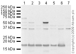 Western blot - Anti-Rab25 antibody (ab45855)All lanes : Anti-Rab25 antibody (ab45855) at 1 µg/ml
Lane 1 : HeLa (Human epithelial carcinoma cell line) Whole Cell Lysate
Lane 2 : HepG2 (Human hepatocellular liver carcinoma cell line) Whole Cell Lysate
Lane 3 : Y79 (Human retinoblastoma cell line) Whole Cell Lysate
Lane 4 :SW480 whole cell lysate (ab3957)
Lane 5 : Rat Thymus Tissue Lysate
Lane 6 : Jurkat (Human T cell lymphoblast-like cell line) Whole Cell Lysate
Lane 7 : Human breast tumor tissue lysate - total protein (ab30091)
Lysates/proteins at 10 µg per lane.
Secondary
All lanes : Goat polyclonal to Rabbit IgG - H&L - Pre-Adsorbed (HRP) at 1/3000 dilution
Developed using the ECL technique.
Performed under reducing conditions.
Predicted band size: 23 kDa
Observed band size: 36 kDa why is the actual band size different from the predicted?
Additional bands at: 17 kDa, 50 kDa, 70 kDa. We are unsure as to the identity of these extra bands. -
 Immunocytochemistry - Anti-Rab25 antibody (ab45855)ICC/IF image of ab45855 stained HeLa cells. The cells were 100% methanol fixed (5 min) and then incubated in 1%BSA / 10% normal goat serum / 0.3M glycine in 0.1% PBS-Tween for 1h to permeabilise the cells and block non-specific protein-protein interactions. The cells were then incubated with the antibody (ab45855, 5µg/ml) overnight at +4°C. The secondary antibody (green) was Alexa Fluor® 488 goat anti-rabbit IgG (H+L) used at a 1/1000 dilution for 1h. Alexa Fluor® 594 WGA was used to label plasma membranes (red) at a 1/200 dilution for 1h. DAPI was used to stain the cell nuclei (blue) at a concentration of 1.43µM. This antibody also gave a positive result in 100% methanol fixed (5 min) Hek293 cells at 5µg/ml, and in 4% PFA fixed (10 min) HeLa and MCF7 cells at 5µg/ml.
Protocols
Datasheets and documents
References (0)
ab45855 has not yet been referenced specifically in any publications.
Images
-
Western blot - Anti-Rab25 antibody (ab45855)All lanes : Anti-Rab25 antibody (ab45855) at 1 µg/ml
Lane 1 : MDA-MB-361 (Human breast adenocarcinoma cell line) Whole Cell Lysate
Lane 2 : MCF7 (Human breast adenocarcinoma cell line) Whole Cell Lysate
Lane 3 : T47D (Human ductal breast epithelial tumor cell line) Whole Cell Lysate
Lysates/proteins at 10 µg per lane.
Secondary
All lanes : Goat polyclonal to Rabbit IgG - H&L - Pre-Adsorbed (HRP) at 1/3000 dilution
Developed using the ECL technique.
Performed under reducing conditions.
Predicted band size: 23 kDa
Observed band size: 32 kDa why is the actual band size different from the predicted?
Additional bands at: 16 kDa, 45 kDa, 75 kDa. We are unsure as to the identity of these extra bands.
-
Western blot - Anti-Rab25 antibody (ab45855)All lanes : Anti-Rab25 antibody (ab45855) at 1 µg/ml
Lane 1 : HeLa (Human epithelial carcinoma cell line) Whole Cell Lysate
Lane 2 : HepG2 (Human hepatocellular liver carcinoma cell line) Whole Cell Lysate
Lane 3 : Y79 (Human retinoblastoma cell line) Whole Cell Lysate
Lane 4 :SW480 whole cell lysate (ab3957)
Lane 5 : Rat Thymus Tissue Lysate
Lane 6 : Jurkat (Human T cell lymphoblast-like cell line) Whole Cell Lysate
Lane 7 : Human breast tumor tissue lysate - total protein (ab30091)
Lysates/proteins at 10 µg per lane.
Secondary
All lanes : Goat polyclonal to Rabbit IgG - H&L - Pre-Adsorbed (HRP) at 1/3000 dilution
Developed using the ECL technique.
Performed under reducing conditions.
Predicted band size: 23 kDa
Observed band size: 36 kDa why is the actual band size different from the predicted?
Additional bands at: 17 kDa, 50 kDa, 70 kDa. We are unsure as to the identity of these extra bands. -
Immunocytochemistry - Anti-Rab25 antibody (ab45855)ICC/IF image of ab45855 stained HeLa cells. The cells were 100% methanol fixed (5 min) and then incubated in 1%BSA / 10% normal goat serum / 0.3M glycine in 0.1% PBS-Tween for 1h to permeabilise the cells and block non-specific protein-protein interactions. The cells were then incubated with the antibody (ab45855, 5µg/ml) overnight at +4°C. The secondary antibody (green) was Alexa Fluor® 488 goat anti-rabbit IgG (H+L) used at a 1/1000 dilution for 1h. Alexa Fluor® 594 WGA was used to label plasma membranes (red) at a 1/200 dilution for 1h. DAPI was used to stain the cell nuclei (blue) at a concentration of 1.43µM. This antibody also gave a positive result in 100% methanol fixed (5 min) Hek293 cells at 5µg/ml, and in 4% PFA fixed (10 min) HeLa and MCF7 cells at 5µg/ml.


