Anti-Nuclear Receptor Corepressor NCoR antibody (ab3482)
")
Key features and details
- Rabbit polyclonal to Nuclear Receptor Corepressor NCoR
- Suitable for: IHC-P, ICC/IF
- Reacts with: Mouse, Human
- Isotype: IgG
Overview
-
Product name
Anti-Nuclear Receptor Corepressor NCoR antibody
See all Nuclear Receptor Corepressor NCoR primary antibodies -
Description
Rabbit polyclonal to Nuclear Receptor Corepressor NCoR -
Host species
Rabbit -
Tested applications
Suitable for: IHC-P, ICC/IFmore details -
Species reactivity
Reacts with: Mouse, Human
Predicted to work with: Rat, Xenopus laevis, Xenopus tropicalis
-
Immunogen
Synthetic peptide corresponding to Mouse Nuclear Receptor Corepressor NCoR aa 2427-2443.
Sequence:PAPLLSAQYETLSDSDD
(Peptide available asab4997)


Properties
-
Form
Liquid -
Storage instructions
Shipped at 4°C. Store at +4°C short term (1-2 weeks). Upon delivery aliquot. Store at -20°C. Avoid freeze / thaw cycle. -
Storage buffer
Preservative: 0.05% Sodium azide
Constituents: 99% PBS, 0.1% BSA -
 Concentration information loading...
 -
Purity
Immunogen affinity purified -
Clonality
Polyclonal -
Isotype
IgG -
Research areas
- Signal Transduction
- Signaling Pathway
- Nuclear Signaling
- Nuclear Hormone Receptors
- Co-activators/co-repressors
- Epigenetics and Nuclear Signaling
- Nuclear Signaling Pathways
- Nuclear Receptors
- Co-activators/co-repressors
- Epigenetics and Nuclear Signaling
- Chromatin Modifying Enzymes
- Acetylation
- HDACs
- Class II / Hda1 Class
Images
-
Immunohistochemistry (Formalin/PFA-fixed paraffin-embedded sections) - Anti-Nuclear Receptor Corepressor NCoR antibody - ChIP Grade (ab3482)
ab3482 labelling Nuclear Receptor Corepressor NCoR in the nucleus of Mouse breast tissue (right) compared with a negative control (left) by Immunohistochemistry (formalin/PFA-fixed paraffin-embedded sections). To expose target proteins, antigen retrieval method was performed using 10mM sodium citrate (pH 6.0), microwaved for 8-15 min. Tissue was blocked in 3% H2O2-methanol for 15 min at room temperature, then incubated with primary antibody (1:200 in 3% BSA-PBS) overnight at 4°C. A HRP-conjugated anti-rabbit was used as the secondary antibody, followed by colorimetric detection using a DAB kit. Tissues were counterstained with hematoxylin and dehydrated with ethanol and xylene to prep for mounting.
-
 Immunohistochemistry (Formalin/PFA-fixed paraffin-embedded sections) - Anti-Nuclear Receptor Corepressor NCoR antibody - ChIP Grade (ab3482)
ab3482 labelling Nuclear Receptor Corepressor NCoR in the nucleus of Human breast carcinoma (right) compared with a negative control (left) by Immunohistochemistry (formalin/PFA-fixed paraffin-embedded sections). To expose target proteins, antigen retrieval method was performed using 10mM sodium citrate (pH 6.0), microwaved for 8-15 min. Tissue was blocked in 3% H2O2-methanol for 15 min at room temperature, then incubated with primary antibody (1:200 in 3% BSA-PBS) overnight at 4°C. A HRP-conjugated anti-rabbit was used as the secondary antibody, followed by colorimetric detection using a DAB kit. Tissues were counterstained with hematoxylin and dehydrated with ethanol and xylene to prep for mounting.
-
 Immunocytochemistry/ Immunofluorescence - Anti-Nuclear Receptor Corepressor NCoR antibody - ChIP Grade (ab3482)
ab3482 labelling Nuclear Receptor Corepressor NCoR (green) in the nucleus and cytoplasm of MCF-7 cells by Immunocytochemistry/Immunofluorescence. Formalin-fixed cells were permeabilized with 0.1% Triton X-100 in TBS for 5-10 minutes and blocked with 3% BSA-PBS for 30 minutes at room temperature. Cells were incubated with the primary antibody (1:200 in 3% BSA-PBS) overnight at 4 ºC. A DyLight-conjugated anti-rabbit was used as the secondary antibody. Red (phalloidin) - F-actin, Blue - nuclei. Images were taken at a magnification of 60x.
-
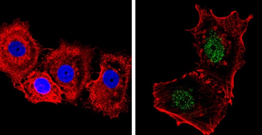
 Immunocytochemistry/ Immunofluorescence - Anti-Nuclear Receptor Corepressor NCoR antibody - ChIP Grade (ab3482)
ab3482 labelling Nuclear Receptor Corepressor NCoR (green) in the nucleus and cytoplasm of HeLa cells by Immunocytochemistry/Immunofluorescence. Formalin-fixed cells were permeabilized with 0.1% Triton X-100 in TBS for 5-10 minutes and blocked with 3% BSA-PBS for 30 minutes at room temperature. Cells were incubated with the primary antibody (1:200 in 3% BSA-PBS) overnight at 4 ºC. A DyLight-conjugated anti-rabbit was used as the secondary antibody. Red (phalloidin) - F-actin, Blue - nuclei. Images were taken at a magnification of 60x.


