Anti-Neuroserpin antibody (ab33077)
Key features and details
- Rabbit polyclonal to Neuroserpin
- Suitable for: WB, ICC/IF
- Reacts with: Mouse, Rat
- Isotype: IgG
Overview
-
Product name
Anti-Neuroserpin antibody
See all Neuroserpin primary antibodies -
Description
Rabbit polyclonal to Neuroserpin -
Host species
Rabbit -
Tested applications
Suitable for: WB, ICC/IFmore details -
Species reactivity
Reacts with: Mouse, Rat
Predicted to work with: Human
-
Immunogen
Synthetic peptide corresponding to Rat Neuroserpin aa 350 to the C-terminus (C terminal) conjugated to keyhole limpet haemocyanin.
(Peptide available asab36619)
Properties
-
Form
Liquid -
Storage instructions
Shipped at 4°C. Store at +4°C short term (1-2 weeks). Upon delivery aliquot. Store at -20°C or -80°C. Avoid freeze / thaw cycle. -
Storage buffer
pH: 7.40
Preservative: 0.02% Sodium azide
Constituent: PBS
Batches of this product that have a concentration Concentration information loading...
Purity
Immunogen affinity purifiedClonality
PolyclonalIsotype
IgGResearch areas
Associated products
-
Compatible Secondaries
-
Isotype control
-
Positive Controls
-
Recombinant Protein
Applications
Our Abpromise guarantee covers the use of ab33077 in the following tested applications.
The application notes include recommended starting dilutions; optimal dilutions/concentrations should be determined by the end user.
Application Abreviews Notes WB ICC/IF Application notesICC/IF: Use at a concentration of 1 µg/ml.
IHC-FoFr: Use at an assay dependent concentration.
WB: 1/250. Detects bands of approximately 46 and 55 kDa (predicted molecular weight: 46 kDa).
Not yet tested in other applications.
Optimal dilutions/concentrations should be determined by the end user.Target
-
Function
Serine protease inhibitor that inhibits plasminogen activators and plasmin but not thrombin. May be involved in the formation or reorganization of synaptic connections as well as for synaptic plasticity in the adult nervous system. May protect neurons from cell damage by tissue-type plasminogen activator. -
Tissue specificity
Predominantly expressed in the brain. -
Involvement in disease
Defects in SERPINI1 are the cause of familial encephalopathy with neuroserpin inclusion bodies (FEN1B) [MIM:604218]. FEN1B is characterized clinically as an autosomal dominantly inherited dementia, histologically by unique neuronal inclusion bodies and biochemically by polymers of neuroserpin. -
Sequence similarities
Belongs to the serpin family. -
Cellular localization
Secreted. - Information by UniProt
-
Database links
- Entrez Gene: 5274 Human
- Entrez Gene: 20713 Mouse
- Entrez Gene: 116459 Rat
- Omim: 602445 Human
- SwissProt: Q99574 Human
- SwissProt: O35684 Mouse
- SwissProt: Q9JLD2 Rat
- Unigene: 478153 Human
see all -
Alternative names
- DKFZp781N13156 antibody
- Neuroserpin antibody
- NEUS_HUMAN antibody
see all
Images
-
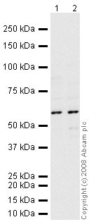
All lanes : Anti-Neuroserpin antibody (ab33077) at 1/250 dilution
Lane 1 : Brain (Mouse) Tissue Lysate
Lane 2 : Brain (Rat) Tissue Lysate
Lysates/proteins at 10 µg per lane.
Secondary
All lanes : Goat polyclonal to Rabbit IgG - H&L - Pre-Adsorbed (HRP) at 1/3000 dilution
Performed under reducing conditions.
Predicted band size: 46 kDa
Observed band size: 55 kDa why is the actual band size different from the predicted?
Neuroserpin contains a number of potential glycosylation sites so it is thought that this is the reason it runs at 55 kDa. The band size observed is also comparable to molecular weights seen with other commercially available antibodies to Neuroserpin. -
ICC/IF image of ab33077 stained rat PC12 cells. The cells were methanol fixed (5 min), permabilised in PBS-T (20 min) and incubated with the antibody (ab33077, 1µg/ml) for 1h at room temperature. 1%BSA / 10% normal goat serum / 0.3M glycine was used to block non-specific protein-protein interactions. The secondary antibody (green) was Alexa Fluor® 488 goat anti-rabbit IgG (H+L) used at a 1/1000 dilution for 1h. Alexa Fluor® 594 WGA was used to label plasma membranes (red). DAPI was used to stain the cell nuclei (blue).
-
All lanes : Anti-Neuroserpin antibody (ab33077) at 1 µg/ml
Lane 1 : Brain (Mouse) Tissue Lysate
Lane 2 : Brain (Rat) Tissue Lysate
Lysates/proteins at 10 µg per lane.
Secondary
All lanes : Goat polyclonal to Rabbit IgG - H&L - Pre-Adsorbed (HRP) at 1/3000 dilution
Performed under reducing conditions.
Predicted band size: 46 kDa
Observed band size: 46,55 kDa why is the actual band size different from the predicted?
Additional bands at: 80 kDa. We are unsure as to the identity of these extra bands.
Exposure time: 10 minutes
Protocols
Datasheets and documents
References (5)
ab33077 has been referenced in 5 publications.
- Adorjan I et al. Neuroserpin expression during human brain development and in adult brain revealed by immunohistochemistry and single cell RNA sequencing. J Anat N/A:N/A (2019). PubMed: 30644551
- Li W et al. Neuroprotective effect of neuroserpin in non-tPA-induced intracerebral hemorrhage mouse models. BMC Neurol 17:196 (2017). WB ; Mouse . PubMed: 29115923
- Kondo S et al. Secretory function in subplate neurons during cortical development. Front Neurosci 9:100 (2015). Mouse . PubMed: 25859180
- Roet KC et al. A multilevel screening strategy defines a molecular fingerprint of proregenerative olfactory ensheathing cells and identifies SCARB2, a protein that improves regenerative sprouting of injured sensory spinal axons. J Neurosci 33:11116-35 (2013). PubMed: 23825416
- Wu J et al. Neuroserpin protects neurons from ischemia-induced plasmin-mediated cell death independently of tissue-type plasminogen activator inhibition. Am J Pathol 177:2576-84 (2010). IHC-FoFr ; Mouse . PubMed: 20864675
Images
-
All lanes : Anti-Neuroserpin antibody (ab33077) at 1/250 dilution
Lane 1 : Brain (Mouse) Tissue Lysate
Lane 2 : Brain (Rat) Tissue Lysate
Lysates/proteins at 10 µg per lane.
Secondary
All lanes : Goat polyclonal to Rabbit IgG - H&L - Pre-Adsorbed (HRP) at 1/3000 dilution
Performed under reducing conditions.
Predicted band size: 46 kDa
Observed band size: 55 kDa why is the actual band size different from the predicted?
Neuroserpin contains a number of potential glycosylation sites so it is thought that this is the reason it runs at 55 kDa. The band size observed is also comparable to molecular weights seen with other commercially available antibodies to Neuroserpin. -
ICC/IF image of ab33077 stained rat PC12 cells. The cells were methanol fixed (5 min), permabilised in PBS-T (20 min) and incubated with the antibody (ab33077, 1µg/ml) for 1h at room temperature. 1%BSA / 10% normal goat serum / 0.3M glycine was used to block non-specific protein-protein interactions. The secondary antibody (green) was Alexa Fluor® 488 goat anti-rabbit IgG (H+L) used at a 1/1000 dilution for 1h. Alexa Fluor® 594 WGA was used to label plasma membranes (red). DAPI was used to stain the cell nuclei (blue).
-
All lanes : Anti-Neuroserpin antibody (ab33077) at 1 µg/ml
Lane 1 : Brain (Mouse) Tissue Lysate
Lane 2 : Brain (Rat) Tissue Lysate
Lysates/proteins at 10 µg per lane.
Secondary
All lanes : Goat polyclonal to Rabbit IgG - H&L - Pre-Adsorbed (HRP) at 1/3000 dilution
Performed under reducing conditions.
Predicted band size: 46 kDa
Observed band size: 46,55 kDa why is the actual band size different from the predicted?
Additional bands at: 80 kDa. We are unsure as to the identity of these extra bands.
Exposure time: 10 minutes


