Anti-MATH2/NEUROD6 antibody (ab85824)
Key features and details
- Rabbit polyclonal to MATH2/NEUROD6
- Suitable for: WB, ICC/IF
- Reacts with: Mouse, Rat, Human
- Isotype: IgG
Overview
-
Product name
Anti-MATH2/NEUROD6 antibody
See all MATH2/NEUROD6 primary antibodies -
Description
Rabbit polyclonal to MATH2/NEUROD6 -
Host species
Rabbit -
Tested applications
Suitable for: WB, ICC/IFmore details -
Species reactivity
Reacts with: Mouse, Rat, Human
Predicted to work with: Cow, Monkey
-
Immunogen
Synthetic peptide corresponding to Human MATH2/NEUROD6 aa 300 to the C-terminus (C terminal) conjugated to keyhole limpet haemocyanin.
(Peptide available asab95917) -
Positive control
- Purchase matching WB positive control:Recombinant Human MATH2/NEUROD6 protein
- This antibody gave a positive signal in the following tissue lysates: Human brain; Mouse brain; Rat brain, as well as the following whole cell lysates: PC12; U87mg; U373 MG Nuclear. ICC/IF: U-87 MG cell line.
-
General notes
Reproducibility is key to advancing scientific discovery and accelerating scientists’ next breakthrough.
Abcam is leading the way with our range of recombinant antibodies, knockout-validated antibodies and knockout cell lines, all of which support improved reproducibility.
We are also planning to innovate the way in which we present recommended applications and species on our product datasheets, so that only applications & species that have been tested in our own labs, our suppliers or by selected trusted collaborators are covered by our Abpromise™ guarantee.
In preparation for this, we have started to update the applications & species that this product is Abpromise guaranteed for.
We are also updating the applications & species that this product has been “predicted to work with,” however this information is not covered by our Abpromise guarantee.
Applications & species from publications and Abreviews that have not been tested in our own labs or in those of our suppliers are not covered by the Abpromise guarantee.
Please check that this product meets your needs before purchasing. If you have any questions, special requirements or concerns, please send us an inquiry and/or contact our Support team ahead of purchase. Recommended alternatives for this product can be found below, as well as customer reviews and Q&As.
Properties
-
Form
Liquid -
Storage instructions
Shipped at 4°C. Store at +4°C short term (1-2 weeks). Upon delivery aliquot. Store at -20°C or -80°C. Avoid freeze / thaw cycle. -
Storage buffer
pH: 7.40
Preservative: 0.02% Sodium azide
Constituent: PBS
Batches of this product that have a concentration Concentration information loading...
Purity
Immunogen affinity purifiedClonality
PolyclonalIsotype
IgGResearch areas
Associated products
-
Compatible Secondaries
-
Isotype control
Applications
Our Abpromise guarantee covers the use of ab85824 in the following tested applications.
The application notes include recommended starting dilutions; optimal dilutions/concentrations should be determined by the end user.
Application Abreviews Notes WB Use a concentration of 1 µg/ml. Detects a band of approximately 40 kDa (predicted molecular weight: 38 kDa). ICC/IF Use a concentration of 5 µg/ml. Target
-
Function
Activates E box-dependent transcription in collaboration with TCF3/E47. May be a trans-acting factor involved in the development and maintenance of the mammalian nervous system. Transactivates the promoter of its own gene. -
Sequence similarities
Contains 1 basic helix-loop-helix (bHLH) domain. -
Cellular localization
Nucleus. - Information by UniProt
-
Database links
- Entrez Gene: 540464 Cow
- Entrez Gene: 63974 Human
- Entrez Gene: 11922 Mouse
- Entrez Gene: 500137 Rat
- Omim: 611513 Human
- SwissProt: Q08DI0 Cow
- SwissProt: Q96NK8 Human
- SwissProt: P48986 Mouse
see all -
Alternative names
- Atoh 2 antibody
- Atoh2 antibody
- Atonal homolog 2 antibody
see all
Images
-
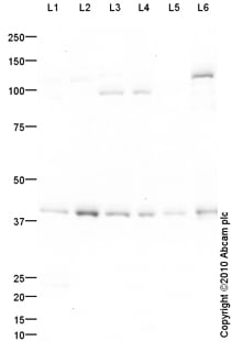
All lanes : Anti-MATH2/NEUROD6 antibody (ab85824) at 1 µg/ml
Lane 1 : PC12 (Rat adrenal pheochromocytoma cell line) Whole Cell Lysate
Lane 2 : U-87 MG (Human glioblastoma astrocytoma) Whole Cell Lysate
Lane 3 : Brain (Mouse) Tissue Lysate
Lane 4 : Brain (Rat) Tissue Lysate
Lane 5 : Human brain tissue lysate - total protein (ab29466)
Lane 6 : U373 MG (Human glioblastoma-astrocytoma, epithelial-like cell line) Nuclear Lysate (ab14902)
Lysates/proteins at 10 µg per lane.
Secondary
All lanes : Goat polyclonal to Rabbit IgG - H&L - Pre-Adsorbed (HRP) at 1/3000 dilution
Developed using the ECL technique.
Performed under reducing conditions.
Predicted band size: 38 kDa
Observed band size: 40 kDa why is the actual band size different from the predicted?
Additional bands at: 100 kDa, 120 kDa. We are unsure as to the identity of these extra bands.
Exposure time: 16 minutes -
ab85824 staining MATH2/NEUROD6 in U-87 MG cells. The cells were fixed with 100% methanol (5 min), permeabilized with 0.1% Triton X-100 for 5 minutes and then blocked with 1% BSA/10% normal goat serum/0.3M glycine in 0.1% PBS-Tween for 1h. The cells were then incubated with the antibody ab85824 at 5µg/ml and ab7291 (Mouse monoclonal to alpha Tubulin - Loading Control) used at a 1/1000 dilution overnight at +4°C. The secondary antibodies were ab150081, Goat Anti-Rabbit IgG H&L (Alexa Fluor® 488) preadsorbed, (pseudo-colored green) and ab150120, Goat polyclonal Secondary Antibody to Mouse IgG - H&L (Alexa Fluor® 594) preadsorbed, (colored red), both used at a 1/1000 dilution for 1 hour at room temperature. DAPI was used to stain the cell nuclei (colored blue) at a concentration of 1.43 µM for 1hour at room temperature.
Protocols
References (5)
ab85824 has been referenced in 5 publications.
- Komura H et al. Alzheimer Aß Assemblies Accumulate in Excitatory Neurons upon Proteasome Inhibition and Kill Nearby NAKa3 Neurons by Secretion. iScience 13:452-477 (2019). PubMed: 30827871
- Luna E et al. Differential a-synuclein expression contributes to selective vulnerability of hippocampal neuron subpopulations to fibril-induced toxicity. Acta Neuropathol 135:855-875 (2018). PubMed: 29502200
- Sullivan CS et al. Perineuronal Net Protein Neurocan Inhibits NCAM/EphA3 Repellent Signaling in GABAergic Interneurons. Sci Rep 8:6143 (2018). PubMed: 29670169
- Tachibana N et al. Pten Regulates Retinal Amacrine Cell Number by Modulating Akt, Tgfß, and Erk Signaling. J Neurosci 36:9454-71 (2016). PubMed: 27605619
- Klisch TJ et al. In vivo Atoh1 targetome reveals how a proneural transcription factor regulates cerebellar development. Proc Natl Acad Sci U S A 108:3288-93 (2011). ICC/IF ; Mouse . PubMed: 21300888
Images
-
All lanes : Anti-MATH2/NEUROD6 antibody (ab85824) at 1 µg/ml
Lane 1 : PC12 (Rat adrenal pheochromocytoma cell line) Whole Cell Lysate
Lane 2 : U-87 MG (Human glioblastoma astrocytoma) Whole Cell Lysate
Lane 3 : Brain (Mouse) Tissue Lysate
Lane 4 : Brain (Rat) Tissue Lysate
Lane 5 : Human brain tissue lysate - total protein (ab29466)
Lane 6 : U373 MG (Human glioblastoma-astrocytoma, epithelial-like cell line) Nuclear Lysate (ab14902)
Lysates/proteins at 10 µg per lane.
Secondary
All lanes : Goat polyclonal to Rabbit IgG - H&L - Pre-Adsorbed (HRP) at 1/3000 dilution
Developed using the ECL technique.
Performed under reducing conditions.
Predicted band size: 38 kDa
Observed band size: 40 kDa why is the actual band size different from the predicted?
Additional bands at: 100 kDa, 120 kDa. We are unsure as to the identity of these extra bands.
Exposure time: 16 minutes
-
ab85824 staining MATH2/NEUROD6 in U-87 MG cells. The cells were fixed with 100% methanol (5 min), permeabilized with 0.1% Triton X-100 for 5 minutes and then blocked with 1% BSA/10% normal goat serum/0.3M glycine in 0.1% PBS-Tween for 1h. The cells were then incubated with the antibody ab85824 at 5µg/ml and ab7291 (Mouse monoclonal to alpha Tubulin - Loading Control) used at a 1/1000 dilution overnight at +4°C. The secondary antibodies were ab150081, Goat Anti-Rabbit IgG H&L (Alexa Fluor® 488) preadsorbed, (pseudo-colored green) and ab150120, Goat polyclonal Secondary Antibody to Mouse IgG - H&L (Alexa Fluor® 594) preadsorbed, (colored red), both used at a 1/1000 dilution for 1 hour at room temperature. DAPI was used to stain the cell nuclei (colored blue) at a concentration of 1.43 µM for 1hour at room temperature.


