Anti-LC3B antibody - N-terminal (ab229327)
Key features and details
- Rabbit polyclonal to LC3B - N-terminal
- Suitable for: WB, Flow Cyt, ICC/IF, IP, IHC-P
- Reacts with: Mouse, Rat, Human
- Isotype: IgG
Overview
-
Product name
Anti-LC3B antibody - N-terminal
See all LC3B primary antibodies -
Description
Rabbit polyclonal to LC3B - N-terminal -
Host species
Rabbit -
Tested Applications & Species
See all applications and species dataApplication Species Flow Cyt HumanICC/IF HumanIHC-P MouseIP HumanWB MouseRatHuman -
Immunogen
Synthetic peptide within Human LC3B (N terminal). The exact sequence is proprietary. (Carrier-protein conjugated).
Database link: Q9GZQ8 -
Positive control
- ICC/IF: HeLa cells untreated and treated with 50µM Chloroquine for 24 hr; Hep G2 cells untreated and treated with 3 µM Thapsigargin for 12 hrs. WB: Huh7 whole cell lysate; NTERA-2 cl.D1 [NT2/D1], PC-3, U-87 MG and SK-N-SH whole cell extracts; Mouse and rat brain lysates; Hep G2 cell lysate untreated and treated with 3 µM Thapsigargin; HeLa whole cell extract untreated and treated with 50µM Chloroquine for 24 hr. IHC-P: Mouse brain tissue. IP: U-87 MG whole cell extract. Flow Cyt: HeLa cells.
Properties
-
Form
Liquid -
Storage instructions
Shipped at 4°C. Store at +4°C short term (1-2 weeks). Upon delivery aliquot. Store at -20°C long term. Avoid freeze / thaw cycle. -
Storage buffer
pH: 7.00
Preservative: 0.025% Proclin 300
Constituents: 78% PBS, 1% BSA, 20% Glycerol (glycerin, glycerine) -
 Concentration information loading...
 -
Purity
Immunogen affinity purified -
Clonality
Polyclonal -
Isotype
IgG -
Research areas
Images
-
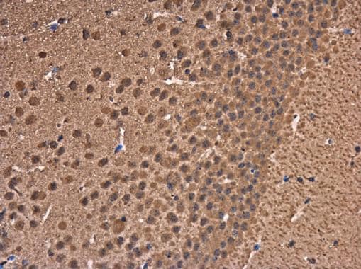
 Immunohistochemistry (Formalin/PFA-fixed paraffin-embedded sections) - Anti-LC3B antibody - N-terminal (ab229327)
Paraffin-embedded mouse brain tissue stained for LC3B using ab229327 at 1/500 dilution in immunohistochemical analysis.
-
All lanes : Anti-LC3B antibody - N-terminal (ab229327) at 1/1500 dilution
Lane 1 : Huh7 whole cell lysate (untreated)
Lane 2 : Huh7 whole cell lysate (3 µM Thapsigargin treatment for 12 hr)
Lysates/proteins at 20 µg per lane.
Secondary
All lanes : HRP-conjugated anti-rabbit IgG
Predicted band size: 15 kDa
-
4% paraformaldehyde-fixed HeLa (human epithelial cell line from cervix adenocarcinoma) cells, mock treated (left panel) or treated with 50 μM Chloroquine for 24 hr (right panel), stained for LC3B (green) using ab229327 at 1/2000 dilution in ICC/IF.
Red: alpha Tubulin, a cytoskeleton marker, stained by alpha Tubulin antibody at 1/1000 dilution.
Blue: Hoechst 33342 staining.
-
All lanes : Anti-LC3B antibody - N-terminal (ab229327) at 1/1500 dilution
Lane 1 : Huh7 whole cell lysate (un-infected)
Lane 2 : Huh7 whole cell lysate (HCV-infected)
Lysates/proteins at 20 µg per lane.
Secondary
All lanes : HRP-conjugated anti-rabbit IgG
Predicted band size: 15 kDa
-
All lanes : Anti-LC3B antibody - N-terminal (ab229327) at 1/1000 dilution
Lane 1 : NTERA-2 cl.D1 [NT2/D1] (human malignant pluripotent embryonic carcinoma cell line) whole cell extract
Lane 2 : PC-3 (human prostate adenocarcinoma cell line) whole cell extract
Lane 3 : U-87 MG (human glioblastoma-astrocytoma epithelial cell line) whole cell extract
Lane 4 : SK-N-SH (human neuroblastoma cell line) whole cell extract
Lysates/proteins at 30 µg per lane.
Secondary
All lanes : HRP-conjugated anti-rabbit IgG
Predicted band size: 15 kDa15% SDS-PAGE gel.
-
Ice-cold methanol-fixed, ice-cold acetone permeabilized HepG2 (human liver hepatocellular carcinoma cell line) cells, mock treated (left panels) or treated with with 3 μM Thapsigargin for 12 hrs (right panels), stained for LC3B (green) using ab229327 at 1/500 dilution in ICC/IF.
Blue: Hoechst 33342 staining.
-
LC3B was immunoprecipitated from U-87 MG (human glioblastoma-astrocytoma epithelial cell line) whole cell extract (1 mg for IP, 20% of IP loaded) with 5 µg ab229327. Western blot was performed from the immunoprecipitate using ab229327. Anti-Rabbit IgG was used as a secondary reagent.
Lane 1: U-87 MG whole cell extract.
Lane 2: Control IgG IP in U-87 MG whole cell extract.
Lane 3: ab229327 IP in U-87 MG whole cell extract.
-
Flow cytometric analysis of 4% paraformaldehyde-fixed HeLa (human epithelial cell line from cervix adenocarcinoma) cell line labeling LC3B with ab229327 at 1/100 dilution (blue) compared with an unlabelled sample (brown).
Acquisition of >20,000 events were collected using Argon ion laser (488nm) and 525/30 bandpass filter.
-
4% paraformaldehyde-fixed HeLa (human epithelial cell line from cervix adenocarcinoma) cells, mock treated (left panels) or treated with 50 μM Chloroquine for 24 hr (right panels), stained for LC3B (green) using ab229327 at 1/200 dilution in ICC/IF.
Red: phalloidin, a cytoskeleton marker, at 1/50 dilution.
Blue: Hoechst 33342 staining.
-
Anti-LC3B antibody - N-terminal (ab229327) at 1/1000 dilution + Mouse brain lysate at 50 µg
Secondary
HRP-conjugated anti-rabbit IgG
Predicted band size: 15 kDa15% SDS-PAGE gel.
-
Anti-LC3B antibody - N-terminal (ab229327) at 1/1000 dilution + Rat brain lysate at 50 µg
Secondary
HRP-conjugated anti-rabbit IgG
Predicted band size: 15 kDa15% SDS-PAGE gel.
-
All lanes : Anti-LC3B antibody - N-terminal (ab229327) at 1/1000 dilution
Lane 1 : Untreated HepG2 (human liver hepatocellular carcinoma cell line) whole cell lysate
Lane 2 : HepG2 whole cell lysate treated with 3 µM thapsigargin for 12 hours
Lysates/proteins at 30 µg per lane.
Secondary
All lanes : HRP-conjugated anti-rabbit IgG
Predicted band size: 15 kDaLower panel: Beta-actin antibody at 1/20000 dilution.
15% SDS-PAGE gel.
-
All lanes : Anti-LC3B antibody - N-terminal (ab229327) at 1/1000 dilution
Lane 1 : Untreated HepG2 (human liver hepatocellular carcinoma cell line) whole cell extract
Lane 2 : HepG2 whole cell extract treated with 3 µM thapsigargin for 16 hours
Lane 3 : HepG2 whole cell extract treated with 3 µM thapsigargin for 24 hours
Lysates/proteins at 30 µg per lane.
Secondary
All lanes : HRP-conjugated anti-rabbit IgG
Predicted band size: 15 kDa15% SDS-PAGE gel.
-
All lanes : Anti-LC3B antibody - N-terminal (ab229327) at 1/2500 dilution
Lane 1 : Untreated HeLa (human epithelial cell line from cervix adenocarcinoma) whole cell extract
Lane 2 : HeLa whole cell extract treated with 50µM Chloroquine for 24 hours
Lysates/proteins at 30 µg per lane.
Secondary
All lanes : HRP-conjugated anti-rabbit IgG
Predicted band size: 15 kDa15% SDS-PAGE gel.


