Anti-LATS2 antibody (ab110780)
Key features and details
- Rabbit polyclonal to LATS2
- Suitable for: IHC-P, WB
- Reacts with: Human
- Isotype: IgG
Overview
-
Product name
Anti-LATS2 antibody
See all LATS2 primary antibodies -
Description
Rabbit polyclonal to LATS2 -
Host species
Rabbit -
Tested Applications & Species
See all applications and species dataApplication Species IHC-P HumanWB Human -
Immunogen
Synthetic peptide. This information is proprietary to Abcam and/or its suppliers.
-
Positive control
- This antibody gave a positive signal in Human Ovary and Human Testis tissue lysates as well as the following whole cell lysates: HeLa; HepG2; MCF7. It also gave a positive signal in FFPE human heart tissue sections.
Properties
-
Form
Liquid -
Storage instructions
Shipped at 4°C. Store at +4°C short term (1-2 weeks). Upon delivery aliquot. Store at -20°C or -80°C. Avoid freeze / thaw cycle. -
Storage buffer
pH: 7.40
Preservative: 0.02% Sodium azide
Constituent: PBS
Batches of this product that have a concentration Concentration information loading...
Purity
Immunogen affinity purifiedClonality
PolyclonalIsotype
IgGResearch areas
Associated products
-
Compatible Secondaries
-
Isotype control
-
Recombinant Protein
Applications
The Abpromise guarantee
Our Abpromise guarantee covers the use of ab110780 in the following tested applications.
The application notes include recommended starting dilutions; optimal dilutions/concentrations should be determined by the end user.
GuaranteedTested applications are guaranteed to work and covered by our Abpromise guarantee.
PredictedPredicted to work for this combination of applications and species but not guaranteed.
IncompatibleDoes not work for this combination of applications and species.
Application Species IHC-P HumanWB HumanAll applications ChimpanzeeGorillaOrangutanApplication Abreviews Notes IHC-P Use a concentration of 10 µg/ml. Perform heat mediated antigen retrieval before commencing with IHC staining protocol.WB Use a concentration of 1 µg/ml. Detects a band of approximately 120 kDa (predicted molecular weight: 120 kDa).Notes IHC-P
Use a concentration of 10 µg/ml. Perform heat mediated antigen retrieval before commencing with IHC staining protocol.WB
Use a concentration of 1 µg/ml. Detects a band of approximately 120 kDa (predicted molecular weight: 120 kDa).Target
-
Function
Negative regulator of YAP1 in the Hippo signaling pathway that plays a pivotal role in organ size control and tumor suppression by restricting proliferation and promoting apoptosis. The core of this pathway is composed of a kinase cascade wherein MST1/MST2, in complex with its regulatory protein SAV1, phosphorylates and activates LATS1/2 in complex with its regulatory protein MOB1, which in turn phosphorylates and inactivates YAP1 oncoprotein and WWTR1/TAZ. Phosphorylation of YAP1 by LATS2 inhibits its translocation into the nucleus to regulate cellular genes important for cell proliferation, cell death, and cell migration. Acts as a tumor suppressor which plays a critical role in centrosome duplication, maintenance of mitotic fidelity and genomic stability. Negatively regulates G1/S transition by down-regulating cyclin E/CDK2 kinase activity. Negative regulator of the androgen receptor. -
Tissue specificity
Expressed at high levels in heart and skeletal muscle and at lower levels in all other tissues examined. -
Sequence similarities
Belongs to the protein kinase superfamily. AGC Ser/Thr protein kinase family.
Contains 1 AGC-kinase C-terminal domain.
Contains 1 protein kinase domain.
Contains 1 UBA domain. -
Post-translational
modificationsAutophosphorylated and phosphorylated during M-phase and the G1/S-phase of the cell cycle. Phosphorylated and activated by STK3. -
Cellular localization
Cytoplasm > cytoskeleton > centrosome. Cytoplasm. Cytoplasm > cytoskeleton > spindle pole. Co-localizes with STK6 at the centrosomes during interphase, early prophase and cytokinesis. Migrates to the spindle poles during mitosis, and to the midbody during cytokinesis. - Information by UniProt
-
Database links
- Entrez Gene: 26524 Human
- Omim: 604861 Human
- SwissProt: Q9NRM7 Human
- Unigene: 78960 Human
-
Alternative names
- FLJ13161 antibody
- Kinase phosphorylated during mitosis protein antibody
- KPM antibody
see all
Images
-
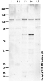
All lanes : Anti-LATS2 antibody (ab110780) at 1 µg/ml
Lane 1 : Human ovary tissue lysate - total protein (ab30222)
Lane 2 : Human testis tissue lysate - total protein (ab30257)
Lane 3 : HeLa (Human epithelial carcinoma cell line) Whole Cell Lysate
Lane 4 : HepG2 (Human hepatocellular liver carcinoma cell line) Whole Cell Lysate
Lane 5 : MCF7 (Human breast adenocarcinoma cell line) Whole Cell Lysate
Lysates/proteins at 10 µg per lane.
Secondary
All lanes : Goat Anti-Rabbit IgG H&L (HRP) preadsorbed (ab97080) at 1/5000 dilution
Developed using the ECL technique.
Performed under reducing conditions.
Predicted band size: 120 kDa
Observed band size: 120 kDa
Additional bands at: 65 kDa, 80 kDa. We are unsure as to the identity of these extra bands.
Exposure time: 20 minutes -
 Immunohistochemistry (Formalin/PFA-fixed paraffin-embedded sections) - Anti-LATS2 antibody (ab110780)IHC image of ab110780 staining in human heart formalin fixed paraffin embedded tissue section, performed on a Leica BondTM system using the standard protocol F. The section was pre-treated using heat mediated antigen retrieval with sodium citrate buffer (pH6, epitope retrieval solution 1) for 20 mins. The section was then incubated with ab110780, 10µg/ml, for 15 mins at room temperature and detected using an HRP conjugated compact polymer system. DAB was used as the chromogen. The section was then counterstained with haematoxylin and mounted with DPX.
For other IHC staining systems (automated and non-automated) customers should optimize variable parameters such as antigen retrieval conditions, primary antibody concentration and antibody incubation times.
Protocols
Datasheets and documents
References (6)
ab110780 has been referenced in 6 publications.
- Wu X et al. RNA Binding Protein RNPC1 Suppresses the Stemness of Human Endometrial Cancer Cells via Stabilizing MST1/2 mRNA. Med Sci Monit 26:e921389 (2020). PubMed: 32088727
- Guo C et al. LATS2 inhibits cell proliferation and metastasis through the Hippo signaling pathway in glioma. Oncol Rep 41:2753-2761 (2019). PubMed: 30896861
- Sun L et al. MicroRNA-744 promotes carcinogenesis in osteosarcoma through targeting LATS2. Oncol Lett 18:2523-2529 (2019). PubMed: 31452740
- Montavon C et al. Outcome in serous ovarian cancer is not associated with LATS expression. J Cancer Res Clin Oncol 145:2737-2749 (2019). PubMed: 31586262
- Li Y et al. Long noncoding RNA DDX11-AS1 epigenetically represses LATS2 by interacting with EZH2 and DNMT1 in hepatocellular carcinoma. Biochem Biophys Res Commun 514:1051-1057 (2019). PubMed: 31097223
- Sadaf et al. Hypermethylated LATS2 gene with decreased expression in female breast cancer: A case control study from North India. Gene 676:156-163 (2018). PubMed: 30010037
Images
-
All lanes : Anti-LATS2 antibody (ab110780) at 1 µg/ml
Lane 1 : Human ovary tissue lysate - total protein (ab30222)
Lane 2 : Human testis tissue lysate - total protein (ab30257)
Lane 3 : HeLa (Human epithelial carcinoma cell line) Whole Cell Lysate
Lane 4 : HepG2 (Human hepatocellular liver carcinoma cell line) Whole Cell Lysate
Lane 5 : MCF7 (Human breast adenocarcinoma cell line) Whole Cell Lysate
Lysates/proteins at 10 µg per lane.
Secondary
All lanes : Goat Anti-Rabbit IgG H&L (HRP) preadsorbed (ab97080) at 1/5000 dilution
Developed using the ECL technique.
Performed under reducing conditions.
Predicted band size: 120 kDa
Observed band size: 120 kDa
Additional bands at: 65 kDa, 80 kDa. We are unsure as to the identity of these extra bands.
Exposure time: 20 minutes
-
 Immunohistochemistry (Formalin/PFA-fixed paraffin-embedded sections) - Anti-LATS2 antibody (ab110780)IHC image of ab110780 staining in human heart formalin fixed paraffin embedded tissue section, performed on a Leica BondTM system using the standard protocol F. The section was pre-treated using heat mediated antigen retrieval with sodium citrate buffer (pH6, epitope retrieval solution 1) for 20 mins. The section was then incubated with ab110780, 10µg/ml, for 15 mins at room temperature and detected using an HRP conjugated compact polymer system. DAB was used as the chromogen. The section was then counterstained with haematoxylin and mounted with DPX.
For other IHC staining systems (automated and non-automated) customers should optimize variable parameters such as antigen retrieval conditions, primary antibody concentration and antibody incubation times.


