Anti-KCND1 antibody (ab101065)
Key features and details
- Rabbit polyclonal to KCND1
- Suitable for: ICC/IF, WB
- Reacts with: Mouse, Human
- Isotype: IgG
Overview
-
Product name
Anti-KCND1 antibody
See all KCND1 primary antibodies -
Description
Rabbit polyclonal to KCND1 -
Host species
Rabbit -
Tested Applications & Species
See all applications and species dataApplication Species ICC/IF HumanWB Mouse -
Immunogen
Synthetic peptide. This information is proprietary to Abcam and/or its suppliers.
-
Positive control
- This antibody gave a positive signal in mouse pancreas tissue lysate.
Properties
-
Form
Liquid -
Storage instructions
Shipped at 4°C. Store at +4°C short term (1-2 weeks). Upon delivery aliquot. Store at -20°C or -80°C. Avoid freeze / thaw cycle. -
Storage buffer
pH: 7.40
Preservative: 0.02% Sodium azide
Constituent: PBS
Batches of this product that have a concentration Concentration information loading...
Purity
Immunogen affinity purifiedClonality
PolyclonalIsotype
IgGResearch areas
Associated products
-
Compatible Secondaries
-
Isotype control
Applications
The Abpromise guarantee
Our Abpromise guarantee covers the use of ab101065 in the following tested applications.
The application notes include recommended starting dilutions; optimal dilutions/concentrations should be determined by the end user.
GuaranteedTested applications are guaranteed to work and covered by our Abpromise guarantee.
PredictedPredicted to work for this combination of applications and species but not guaranteed.
IncompatibleDoes not work for this combination of applications and species.
Application Species ICC/IF HumanWB MouseApplication Abreviews Notes ICC/IF Use a concentration of 10 µg/ml.WB Use a concentration of 1 µg/ml. Detects a band of approximately 71 kDa (predicted molecular weight: 71 kDa).Notes ICC/IF
Use a concentration of 10 µg/ml.WB
Use a concentration of 1 µg/ml. Detects a band of approximately 71 kDa (predicted molecular weight: 71 kDa).Target
-
Function
Pore-forming (alpha) subunit of voltage-gated rapidly inactivating A-type potassium channels. May contribute to I(To) current in heart and I(Sa) current in neurons. Channel properties are modulated by interactions with other alpha subunits and with regulatory subunits. -
Tissue specificity
Widely expressed. Highly expressed in brain, in particular in cerebellum and thalamus; detected at lower levels in the other parts of the brain. -
Sequence similarities
Belongs to the potassium channel family. D (Shal) (TC 1.A.1.2) subfamily. Kv4.1/KCND1 sub-subfamily. -
Domain
The segment S4 is probably the voltage-sensor and is characterized by a series of positively charged amino acids at every third position. -
Cellular localization
Membrane. - Information by UniProt
-
Database links
- Entrez Gene: 3750 Human
- Entrez Gene: 16506 Mouse
- Omim: 300281 Human
- SwissProt: Q9NSA2 Human
- SwissProt: Q03719 Mouse
- Unigene: 55276 Human
- Unigene: 335968 Mouse
-
Alternative names
- Kcnd1 antibody
- KCND1_HUMAN antibody
- Kv4.1 antibody
see all
Images
-
Anti-KCND1 antibody (ab101065) at 1 µg/ml + Pancreas (Mouse) Tissue Lysate at 10 µg
Secondary
Goat Anti-Rabbit IgG H&L (HRP) preadsorbed (ab97080) at 1/5000 dilution
Developed using the ECL technique.
Performed under reducing conditions.
Predicted band size: 71 kDa
Observed band size: 71 kDa
Exposure time: 16 minutes -
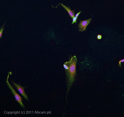
ICC/IF image of ab101065 stained SKNSH cells. The cells were 4% formaldehyde fixed (10 min) and then incubated in 1%BSA / 10% normal goat serum / 0.3M glycine in 0.1% PBS-Tween for 1h to permeabilise the cells and block non-specific protein-protein interactions. The cells were then incubated with the antibody ab101065 at 10µg/ml overnight at +4°C. The secondary antibody (green) was DyLight® 488 goat anti- rabbit (ab96899) IgG (H+L) used at a 1/1000 dilution for 1h. Alexa Fluor® 594 WGA was used to label plasma membranes (red) at a 1/200 dilution for 1h. DAPI was used to stain the cell nuclei (blue) at a concentration of 1.43µM.
Protocols
Datasheets and documents
References (0)
ab101065 has not yet been referenced specifically in any publications.
Images
-
Anti-KCND1 antibody (ab101065) at 1 µg/ml + Pancreas (Mouse) Tissue Lysate at 10 µg
Secondary
Goat Anti-Rabbit IgG H&L (HRP) preadsorbed (ab97080) at 1/5000 dilution
Developed using the ECL technique.
Performed under reducing conditions.
Predicted band size: 71 kDa
Observed band size: 71 kDa
Exposure time: 16 minutes
-
ICC/IF image of ab101065 stained SKNSH cells. The cells were 4% formaldehyde fixed (10 min) and then incubated in 1%BSA / 10% normal goat serum / 0.3M glycine in 0.1% PBS-Tween for 1h to permeabilise the cells and block non-specific protein-protein interactions. The cells were then incubated with the antibody ab101065 at 10µg/ml overnight at +4°C. The secondary antibody (green) was DyLight® 488 goat anti- rabbit (ab96899) IgG (H+L) used at a 1/1000 dilution for 1h. Alexa Fluor® 594 WGA was used to label plasma membranes (red) at a 1/200 dilution for 1h. DAPI was used to stain the cell nuclei (blue) at a concentration of 1.43µM.


