Anti-KAT13D / CLOCK antibody (ab3517)
")
Key features and details
- Rabbit polyclonal to KAT13D / CLOCK
- Suitable for: ICC, IHC-P, WB
- Reacts with: Mouse, Human
- Isotype: IgG
Overview
-
Product name
Anti-KAT13D / CLOCK antibody
See all KAT13D / CLOCK primary antibodies -
Description
Rabbit polyclonal to KAT13D / CLOCK -
Host species
Rabbit -
Tested Applications & Species
See all applications and species dataApplication Species ICC HumanIHC-P MouseHumanWB MouseHuman
-
Immunogen
Synthetic peptide corresponding to Mouse KAT13D/ CLOCK aa 1-100.
-
General notes
The Life Science industry has been in the grips of a reproducibility crisis for a number of years. Abcam is leading the way in addressing the problem with our range of recombinant monoclonal antibodies and knockout edited cell lines for gold-standard validation.
One factor contributing to the crisis is the use of antibodies that are not suitable. This can lead to misleading results and the use of incorrect data informing project assumptions and direction. To help address this challenge, we have introduced an application and species grid on our primary antibody datasheets to make it easy to simplify identification of the right antibody for your needs.
Learn more here.


Properties
-
Form
Liquid -
Storage instructions
Shipped at 4°C. Store at +4°C short term (1-2 weeks). Upon delivery aliquot. Store at -20°C or -80°C. Avoid freeze / thaw cycle. -
Storage buffer
Preservative: 0.05% Sodium azide
Constituents: 0.1% BSA, 99% PBS -
 Concentration information loading...
 -
Purity
Immunogen affinity purified -
Clonality
Polyclonal -
Isotype
IgG -
Research areas
Images
-
Immunocytochemistry - Anti-KAT13D / CLOCK antibody (ab3517)
Immunocytochemistry/immunofluorescence analysis of U251 cells labeling KAT13D/CLOCK (green) with ab3517 at 1/100. Cells were fixed with formalin and permeabilized with 0.1% Triton X-100 in TBS for 5-10 minutes and blcoked with £% BSA in PBS for 30 minutes at room temperature. Cells were incubated with the primary antibody overnight at 4°C. A DyLight-conjugated secondary antibody was used. F-actin (red) was stained with phalloidin and nuclei (blue) were stained with Hoechst or DAPI. 60X magnification. Left - negative control.
-
 Immunohistochemistry (Formalin/PFA-fixed paraffin-embedded sections) - Anti-KAT13D / CLOCK antibody (ab3517)
ab3517 labelling KAT13D in the nucleus and cytoplasm of Human skeletal muscle tissue (right) compared with a negative control (left) by Immunohistochemistry (formalin/PFA-fixed paraffin-embedded sections). To expose target proteins, antigen retrieval method was performed using 10mM sodium citrate (pH 6.0) microwaved for 8-15 min. Following antigen retrieval, tissues were blocked in 3% H2O2-methanol for 15 min at room temperature. Thissue sections were incubated with the primary antibody (1:200 in 3% BSA-PBS) overnight at 4°C. A HRP-conjugated anti-rabbit IgG was used as the secondary antibody, followed by colorimetric detection using a DAB kit. Tissues were counterstained with hematoxylin and dehydrated with ethanol and xylene to prep for mounting.
-
 Immunohistochemistry (Formalin/PFA-fixed paraffin-embedded sections) - Anti-KAT13D / CLOCK antibody (ab3517)
ab3517 labelling KAT13D in the nucleus and cytoplasm of Mouse colon tissue (right) compared with a negative control (left) by Immunohistochemistry (formalin/PFA-fixed paraffin-embedded sections). To expose target proteins, antigen retrieval method was performed using 10mM sodium citrate (pH 6.0) microwaved for 8-15 min. Following antigen retrieval, tissues were blocked in 3% H2O2-methanol for 15 min at room temperature. Thissue sections were incubated with the primary antibody (1:200 in 3% BSA-PBS) overnight at 4°C. A HRP-conjugated anti-rabbit IgG was used as the secondary antibody, followed by colorimetric detection using a DAB kit. Tissues were counterstained with hematoxylin and dehydrated with ethanol and xylene to prep for mounting.
-
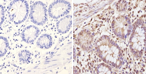 Immunohistochemistry (Formalin/PFA-fixed paraffin-embedded sections) - Anti-KAT13D / CLOCK antibody (ab3517)
ab3517 labelling KAT13D in the nucleus and cytoplasm of Human colon tissue (right) compared with a negative control (left) by Immunohistochemistry (formalin/PFA-fixed paraffin-embedded sections). To expose target proteins, antigen retrieval method was performed using 10mM sodium citrate (pH 6.0) microwaved for 8-15 min. Following antigen retrieval, tissues were blocked in 3% H2O2-methanol for 15 min at room temperature. Thissue sections were incubated with the primary antibody (1:200 in 3% BSA-PBS) overnight at 4°C. A HRP-conjugated anti-rabbit IgG was used as the secondary antibody, followed by colorimetric detection using a DAB kit. Tissues were counterstained with hematoxylin and dehydrated with ethanol and xylene to prep for mounting.
-
 Immunohistochemistry (Formalin/PFA-fixed paraffin-embedded sections) - Anti-KAT13D / CLOCK antibody (ab3517) This image is courtesy of an anonymous Abreviewab3517 staining KAT13D/CLOCK in Mouse skeletal muscle tissue sections by IHC-P (Paraformaldehyde-fixed, paraffin-embedded tissue sections). Tissue was fixed with paraformaldehyde and blocked with 10% serum for 1 hour at 20°C; antigen retrieval was by heat mediation in citrate buffer pH6. Samples were incubated with primary antibody (1/400 in PBS) for 12 hours at 4°C. Undiluted ab64256 was used as the secondary antibody.
 -
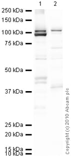
 Western blot - Anti-KAT13D / CLOCK antibody (ab3517)All lanes : Anti-KAT13D / CLOCK antibody (ab3517) at 1/4000 dilution
Lane 1 : HeLa cell lysate
Lane 2 : NIH-3T3 cell lysate
Lysates/proteins at 25 µg per lane.
Predicted band size: 95 kDa
Observed band size: 100 kDa why is the actual band size different from the predicted?
-
 Western blot - Anti-KAT13D / CLOCK antibody (ab3517)All lanes : Anti-KAT13D / CLOCK antibody (ab3517) at 1 µg/ml
Lane 1 : Skeletal Muscle (Mouse) Tissue Lysate
Lane 2 : HeLa (Human epithelial carcinoma cell line) Whole Cell Lysate
Lysates/proteins at 10 µg per lane.
Secondary
All lanes : Goat Anti-Rabbit IgG H&L (HRP) preadsorbed (ab97080) at 1/5000 dilution
Developed using the ECL technique.
Performed under reducing conditions.
Predicted band size: 95 kDa
Observed band size: 95 kDa
Additional bands at: 105 kDa (possible post-translational modification), 38 kDa, 45 kDa. We are unsure as to the identity of these extra bands.
Exposure time: 30 seconds
KAT13D / CLOCK contains a number of potential phosphorylation sites (SwissProt) which may explain the banding pattern observed at a higher molecular weight than predicted.


