Anti-Histone H2A.X antibody - ChIP Grade (ab20669)
")
Key features and details
- Rabbit polyclonal to Histone H2A.X - ChIP Grade
- Suitable for: ChIP, WB, IP, IHC-P
- Knockout validated
- Reacts with: Mouse, Rat, Human, Recombinant fragment
- Isotype: IgG
Overview
-
Product name
Anti-Histone H2A.X antibody - ChIP Grade
See all Histone H2A.X primary antibodies -
Description
Rabbit polyclonal to Histone H2A.X - ChIP Grade -
Host species
Rabbit -
Tested Applications & Species
See all applications and species dataApplication Species ChIP HumanIHC-P HumanIP RatWB MouseHuman
-
Immunogen
Synthetic peptide corresponding to Human Histone H2A.X aa 100 to the C-terminus (C terminal) conjugated to keyhole limpet haemocyanin.
(Peptide available asab15646)
Images
-
Western blot - Anti-Histone H2A.X antibody - ChIP Grade (ab20669)All lanes : Anti-Histone H2A.X antibody - ChIP Grade (ab20669) at 1 µg/ml
Lane 1 : Wild-type HAP1 whole cell lysate
Lane 2 : H2AFX (Histone H2A.X) knockout HAP1 whole cell lysate
Lane 3 : HEK 293 whole cell lysate
Lysates/proteins at 20 µg per lane.
Predicted band size: 15 kDaLanes 1 - 3: Merged signal (red and green). Green - ab20669 observed at 17 kDa. Red - loading control, ab9484, observed at 37 kDa.
ab20669 was shown to recognize Histone H2A.X in wild-type HAP1 cells as signal was lost at the expected MW in H2AFX (Histone H2A.X) knockout cells. Additional cross-reactive bands were observed in the wild-type and knockout cells. Wild-type and H2AFX (Histone H2A.X) knockout samples were subjected to SDS-PAGE. Ab20669 and ab9484 (Mouse anti-GAPDH loading control) were incubated overnight at 4°C at 1 μg/ml and 1/20000 dilution respectively. Blots were developed with Goat anti-Rabbit IgG H&L (IRDye® 800CW) preabsorbed ab216773 and Goat anti-Mouse IgG H&L (IRDye® 680RD) preabsorbed ab216776 secondary antibodies at 1/20000 dilution for 1 hour at room temperature before imaging.
-
 Western blot - Anti-Histone H2A.X antibody - ChIP Grade (ab20669)All lanes : Anti-Histone H2A.X antibody - ChIP Grade (ab20669) at 1 µg/ml
Lane 1 : HeLa (Human epithelial carcinoma cell line) Nuclear Lysate at 10 µg
Lane 2 : HeLa Histone Preparation Nuclear Lysate at 2.5 µg
Lane 3 : Histone H2A Recombinant Protein (negative control) at 0.1 µg
Secondary
All lanes : Goat polyclonal to Rabbit IgG - H&L - Pre-Adsorbed (HRP) (ab65484) at 1/3000 dilution
Developed using the ECL technique.
Performed under reducing conditions.
Predicted band size: 15 kDa
Observed band size: 17 kDa why is the actual band size different from the predicted?
Exposure time: 20 minutes
-
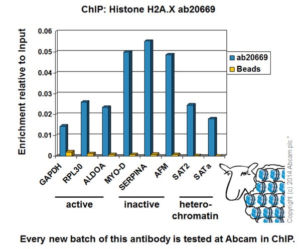 ChIP - Anti-Histone H2A.X antibody - ChIP Grade (ab20669)
Chromatin was prepared from HeLa cells according to the Abcam X-ChIP protocol. Cells were fixed with formaldehyde for 10 minutes. The ChIP was performed with 25µg of chromatin, 2µg of ab20669 (blue), and 20µl of Protein A/G sepharose beads. No antibody was added to the beads control (yellow). The immunoprecipitated DNA was quantified by real time PCR (Taqman approach for active and inactive loci, Sybr green approach for heterochromatic loci). Primers and probes are located in the first kb of the transcribed region.
-
 Immunohistochemistry (Formalin/PFA-fixed paraffin-embedded sections) - Anti-Histone H2A.X antibody - ChIP Grade (ab20669)
IHC image of ab20669 staining Histone H2A.X in human colon formalin fixed paraffin embedded tissue sections, performed on a Leica Bond. The section was pre-treated using heat mediated antigen retrieval with sodium citrate buffer (pH6, epitope retrieval solution 1) for 20 mins. The section was then incubated with ab20669, 0.5µg/ml, for 15 mins at room temperature and detected using an HRP conjugated compact polymer system. DAB was used as the chromogen. The section was then counterstained with haematoxylin and mounted with DPX. No primary antibody was used in the negative control (shown on the inset).
For other IHC staining systems (automated and non-automated) customers should optimize variable parameters such as antigen retrieval conditions, primary antibody concentration and antibody incubation times.
-
 Immunocytochemistry/ Immunofluorescence - Anti-Histone H2A.X antibody - ChIP Grade (ab20669) This image is courtesy of an anonymous Abreview
ab20669 staining Histone H2A.X in Mouse brain neurone cells by ICC/IF (Immunocytochemistry/immunofluorescence). Cells were fixed with paraformaldehyde, permeabilized with 0.3% Triton X-100 and blocked with 5% serum for 2 hours at 22°C. Samples were incubated with primary antibody (1/500 in 2.5% serum + 0.3% Triton X-100 in PBS) for 24 hours at 4°C. ab175651, an Alexa Fluor® 405-conjugated Donkey anti-rabbit IgG polyclonal (1/500) was used as the secondary antibody.
-
 Immunohistochemistry (Formalin/PFA-fixed paraffin-embedded sections) - Anti-Histone H2A.X antibody - ChIP Grade (ab20669)
IHC image of Histone H2A.X staining in Human Lung formalin fixed paraffin embedded tissue section, performed on a Leica Bond™ system using the standard protocol F. The section was pre-treated using heat mediated antigen retrieval with sodium citrate buffer (pH6, epitope retrieval solution 1) for 20 mins. The section was then incubated with ab20669, 0.2 µg/ml, for 15 mins at room temperature and detected using an HRP conjugated compact polymer system. DAB was used as the chromogen. The section was then counterstained with haematoxylin and mounted with DPX.
For other IHC staining systems (automated and non-automated) customers should optimize variable parameters such as antigen retrieval conditions, primary antibody concentration and antibody incubation times.
-
 Immunocytochemistry/ Immunofluorescence - Anti-Histone H2A.X antibody - ChIP Grade (ab20669) This image is courtesy of an Abreview submitted by Dr Kirk McManusab20669 (1/2000) staining Histone H2A.X in Hela cells (green). Cells were fixed in methanol and counter-stained with DAPI in order to highlight the nucleus (red). Please refer to abreview for further experimental details.
 -
 Immunoprecipitation - Anti-Histone H2A.X antibody - ChIP Grade (ab20669)Histone H2A.X was immunoprecipitated using 0.5mg Rat Testis whole tissue extract, 5µg of Rabbit polyclonal to Histone H2A.X and 50µl of protein G magnetic beads (+). No antibody was added to the control (-).
The antibody was incubated under agitation with Protein G beads for 10min, Rat Testis whole tissue extract lysate diluted in RIPA buffer was added to each sample and incubated for a further 10min under agitation.
Proteins were eluted by addition of 40µl SDS loading buffer and incubated for 10min at 70oC; 10µl of each sample was separated on a SDS PAGE gel, transferred to a nitrocellulose membrane, blocked with 5% BSA and probed with ab20669.
Secondary: Clean blot (HRP conjugate) at 1/1000 dilution.
Band: 17kDa: Histone H2A.X. -
 Western blot - Anti-Histone H2A.X antibody - ChIP Grade (ab20669)All lanes : Anti-Histone H2A.X antibody - ChIP Grade (ab20669) at 1 µg/ml
Lane 1 : Lung (Mouse) Tissue Lysate
Lane 2 : Testis (Mouse) Tissue Lysate
Lane 3 : Lung (Rat) Tissue Lysate
Lane 4 : Testis (Rat) Tissue Lysate - normal tissue (ab29388)
Lysates/proteins at 10 µg per lane.
Secondary
All lanes : Goat polyclonal to Rabbit IgG - H&L - Pre-Adsorbed (HRP) at 1/3000 dilution
Developed using the ECL technique.
Performed under reducing conditions.
Predicted band size: 15 kDa
Observed band size: 17 kDa why is the actual band size different from the predicted?
Additional bands at: 55 kDa. We are unsure as to the identity of these extra bands.
Exposure time: 8 minutes


