Anti-Histone H1.2 antibody - ChIP Grade (ab4086)
")
Key features and details
- Rabbit polyclonal to Histone H1.2 - ChIP Grade
- Suitable for: ChIP, WB, IP, ICC/IF, IHC-P
- Knockout validated
- Reacts with: Human
- Isotype: IgG
Overview
-
Product name
Anti-Histone H1.2 antibody - ChIP Grade
See all Histone H1.2 primary antibodies -
Description
Rabbit polyclonal to Histone H1.2 - ChIP Grade -
Host species
Rabbit -
Specificity
This antibody has only been tested on bulk HeLa histones. We expect this antibody to be specific for Histone H1.2 - however we have not tested this specifically on different histone H1 variants. -
Tested Applications & Species
See all applications and species dataApplication Species ChIP HumanICC/IF HumanIHC-P HumanIP HumanWB Human
-
Immunogen
Synthetic peptide corresponding to Human Histone H1.2 aa 1-100 conjugated to keyhole limpet haemocyanin.
(Peptide available asab16936) -
Positive control
- HeLa Histone preparation
-
General notes
Histone H1.2 is one of five known h1 variants (known as H1.1/2/3/4/5 and/or H1.a/b/c/d/e). The H1 variants differ from each other in the amino acid sequence of their N-terminal regions.
Properties
-
Form
Liquid -
Storage instructions
Shipped at 4°C. Store at +4°C short term (1-2 weeks). Upon delivery aliquot. Store at -20°C or -80°C. Avoid freeze / thaw cycle. -
Storage buffer
pH: 7.40
Preservative: 0.02% Sodium azide
Constituent: PBS
Batches of this product that have a concentration Concentration information loading...
Purity
Immunogen affinity purifiedPrimary antibody notes
Histone H1.2 is one of five known h1 variants (known as H1.1/2/3/4/5 and/or H1.a/b/c/d/e). The H1 variants differ from each other in the amino acid sequence of their N-terminal regions.Clonality
PolyclonalIsotype
IgGResearch areas
Associated products
-
ChIP Related Products
-
Compatible Secondaries
-
Immunizing Peptide (Blocking)
-
Isotype control
-
Recombinant Protein
-
Related Products
Applications
The Abpromise guarantee
Our Abpromise guarantee covers the use of ab4086 in the following tested applications.
The application notes include recommended starting dilutions; optimal dilutions/concentrations should be determined by the end user.
GuaranteedTested applications are guaranteed to work and covered by our Abpromise guarantee.
PredictedPredicted to work for this combination of applications and species but not guaranteed.
IncompatibleDoes not work for this combination of applications and species.
Application Species ChIP HumanICC/IF HumanIHC-P HumanIP HumanWB HumanApplication Abreviews Notes ChIP Use 2-6 µg for 25 µg of chromatin.WB (2) 1/500. Detects a band of approximately 25 kDa (predicted molecular weight: 21 kDa).IP Use a concentration of 5 µg/ml.ICC/IF Use a concentration of 1 µg/ml.IHC-P Use a concentration of 1 µg/ml. Perform heat mediated antigen retrieval before commencing with IHC staining protocol.Notes ChIP
Use 2-6 µg for 25 µg of chromatin.WB
1/500. Detects a band of approximately 25 kDa (predicted molecular weight: 21 kDa).IP
Use a concentration of 5 µg/ml.ICC/IF
Use a concentration of 1 µg/ml.IHC-P
Use a concentration of 1 µg/ml. Perform heat mediated antigen retrieval before commencing with IHC staining protocol.Target
-
Function
Histones H1 are necessary for the condensation of nucleosome chains into higher order structures. -
Sequence similarities
Belongs to the histone H1/H5 family.
Contains 1 H15 (linker histone H1/H5 globular) domain. -
Cellular localization
Nucleus. Chromosome. - Information by UniProt
-
Database links
- Entrez Gene: 3006 Human
- Omim: 142710 Human
- SwissProt: P16403 Human
- Unigene: 7644 Human
-
Alternative names
- H1 histone family member 2 antibody
- H1.a antibody
- H12_HUMAN antibody
see all
Images
-
Western blot - Anti-Histone H1.2 antibody - ChIP Grade (ab4086)All lanes : Anti-Histone H1.2 antibody - ChIP Grade (ab4086) at 1/500 dilution
Lane 1 : Wild-type A549 whole cell lysate at 20 µg/ml
Lane 2 : HeLa whole cell lysate at 20 µg/ml
Lane 3 : HIST1H1C knockout A549 whole cell lysate at 20 µg
Predicted band size: 21 kDaLanes 1 - 3: Merged signal (red and green). Green - ab4086 observed at 30 kDa. Red - loading control, ab130007, observed at 130 kDa.
ab4086 was shown to recognize HIST1H1C in wild-type A549 cells as signal was lost at the expected MW in HeLa knockout cells. Additional cross-reactive bands were observed in the wild-type and knockout cells. Wild-type and HeLa knockout samples were subjected to SDS-PAGE. Ab4086 and ab130007 (Mouse anti-Vinculin loading control) were incubated overnight at 4°C at 1/500 dilution and 1/20000 dilution respectively. Blots were developed with Goat anti-Rabbit IgG H&L (IRDye® 800CW) preabsorbed ab216773 and Goat anti-Mouse IgG H&L (IRDye® 680RD) preabsorbed ab216776 secondary antibodies at 1/20000 dilution for 1 hour at room temperature before imaging.
-
 Immunocytochemistry/ Immunofluorescence - Anti-Histone H1.2 antibody - ChIP Grade (ab4086)
ICC/IF image of ab4086 stained human HeLa cells. The cells were PFA fixed (3.7% PFA, 10 min) and incubated with the antibody (ab4086, 1µg/ml) for 1h at room temperature. The secondary antibody (green) was Alexa Fluor® 488 goat anti-rabbit IgG (H+L) used at a 1/1000 dilution for 1h. Image-iTTM FX Signal Enhancer was used as the primary blocking agent, 5% BSA (in TBS-T) was used for all other blocking steps. DAPI was used to stain the cell nuclei (blue). Alexa Fluor® 594 WGA was used to label plasma membranes (red).
-
 Western blot - Anti-Histone H1.2 antibody - ChIP Grade (ab4086)
ab4086 Histone H1.2
The antibody was used at a dilution of 1/500
Lane 1: HeLa Histone (5ug) + ab4086
Lane 2: HeLa Histone (5ug) + ab4086 + 1µ Secondary ab: Goat polyclonal to Rabbit IgG H&L (HRP) Pre-Adsorbed ab7090 (1/5000)
Exposure time: 1 minute
ab4086 Histone H1.2
Expected molecular weight: 21.3 kDa
The antibody was used at a dilution of 1/500
Lane 1: HeLa Histone (5ug) + ab4086
Lane 2: HeLa Histone (5ug) + ab4086 + 1µg/ml of peptide (Histone H1.2) (ab16936)
Secondary ab: Goat polyclonal to Rabbit IgG H&L (HRP) Pre-Adsorbed ab7090 (1/5000)
Exposure time: 1 minute
Expected molecular weight: 21.3 kDa -
 Immunoprecipitation - Anti-Histone H1.2 antibody - ChIP Grade (ab4086)
Histone H1.2 - ChIP Grade was immunoprecipitated using 0.5mg HeLa whole cell extract, 5µg of Rabbit polyclonal to and 50µl of protein G magnetic beads (+). No antibody was added to the control (-).
The antibody was incubated under agitation with Protein G beads for 10min, HeLa whole cell extract lysate diluted in RIPA buffer was added to each sample and incubated for a further 10min under agitation.
Proteins were eluted by addition of 40µl SDS loading buffer and incubated for 10min at 70°C; 10µl of each sample was separated on a SDS PAGE gel, transferred to a nitrocellulose membrane, blocked with 5% BSA and probed with ab4086.
Secondary: Mouse monoclonal [SB62a] Secondary Antibody to Rabbit IgG light chain (HRP) (ab99697).
Band: 21kDa; Histone H1.2 - ChIP Grade
-
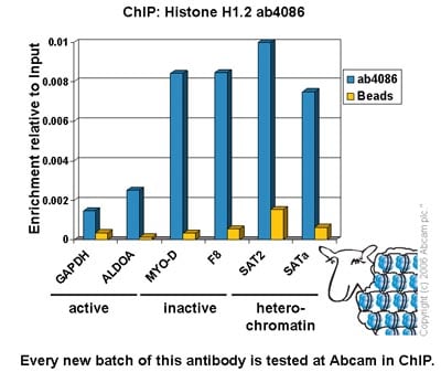 ChIP - Anti-Histone H1.2 antibody - ChIP Grade (ab4086)
Chromatin was prepared from Hela cells according to the Abcam X-ChIP protocol. Cells were fixed with formaldehyde for 10min. The ChIP was performed with 25µg of chromatin, 2µg of ab4086 (blue), and 20µl of Protein A/G sepharose beads. No antibody was added to the beads control (yellow). The immunoprecipitated DNA was quantified by real time PCR (Taqman approach for active and inactive loci, Sybr green approach for heterochromatic loci). Primers and probes are located in the first kb of the transcribed region.
-
 Immunohistochemistry (Formalin/PFA-fixed paraffin-embedded sections) - Anti-Histone H1.2 antibody - ChIP Grade (ab4086)IHC image of Histone H1.2 staining in human breast carcinoma FFPE section, performed on a BondTM system using the standard protocol F. The section was pre-treated using heat mediated antigen retrieval with sodium citrate buffer (pH6, epitope retrieval solution 1) for 20 mins. The section was then incubated with ab4086, 1µg/ml, for 8 mins at room temperature and detected using an HRP conjugated compact polymer system. DAB was used as the chromogen. The section was then counterstained with haematoxylin and mounted with DPX.
Protocols
To our knowledge, customised protocols are not required for this product. Please try the standard protocols listed below and let us know how you get on.
Datasheets and documents
References (16)
ab4086 has been referenced in 16 publications.
- Shimada M et al. Gene-Specific H1 Eviction through a Transcriptional Activator?p300?NAP1?H1 Pathway. Mol Cell 74:268-283.e5 (2019). PubMed: 30902546
- Chiarella E et al. ZNF521 Has an Inhibitory Effect on the Adipogenic Differentiation of Human Adipose-Derived Mesenchymal Stem Cells. Stem Cell Rev N/A:N/A (2018). PubMed: 29938352
- Schauwecker SM et al. Histone H1 and Chromosomal Protein HMGN2 Regulate Prolactin-induced STAT5 Transcription Factor Recruitment and Function in Breast Cancer Cells. J Biol Chem 292:2237-2254 (2017). PubMed: 28035005
- Liao R & Mizzen CA Site-specific regulation of histone H1 phosphorylation in pluripotent cell differentiation. Epigenetics Chromatin 10:29 (2017). PubMed: 28539972
- Munro S et al. Linker Histone H1.2 Directs Genome-wide Chromatin Association of the Retinoblastoma Tumor Suppressor Protein and Facilitates Its Function. Cell Rep 19:2193-2201 (2017). PubMed: 28614707
Images
-
Western blot - Anti-Histone H1.2 antibody - ChIP Grade (ab4086)All lanes : Anti-Histone H1.2 antibody - ChIP Grade (ab4086) at 1/500 dilution
Lane 1 : Wild-type A549 whole cell lysate at 20 µg/ml
Lane 2 : HeLa whole cell lysate at 20 µg/ml
Lane 3 : HIST1H1C knockout A549 whole cell lysate at 20 µg
Predicted band size: 21 kDaLanes 1 - 3: Merged signal (red and green). Green - ab4086 observed at 30 kDa. Red - loading control, ab130007, observed at 130 kDa.
ab4086 was shown to recognize HIST1H1C in wild-type A549 cells as signal was lost at the expected MW in HeLa knockout cells. Additional cross-reactive bands were observed in the wild-type and knockout cells. Wild-type and HeLa knockout samples were subjected to SDS-PAGE. Ab4086 and ab130007 (Mouse anti-Vinculin loading control) were incubated overnight at 4°C at 1/500 dilution and 1/20000 dilution respectively. Blots were developed with Goat anti-Rabbit IgG H&L (IRDye® 800CW) preabsorbed ab216773 and Goat anti-Mouse IgG H&L (IRDye® 680RD) preabsorbed ab216776 secondary antibodies at 1/20000 dilution for 1 hour at room temperature before imaging.
-
Immunocytochemistry/ Immunofluorescence - Anti-Histone H1.2 antibody - ChIP Grade (ab4086)
ICC/IF image of ab4086 stained human HeLa cells. The cells were PFA fixed (3.7% PFA, 10 min) and incubated with the antibody (ab4086, 1µg/ml) for 1h at room temperature. The secondary antibody (green) was Alexa Fluor® 488 goat anti-rabbit IgG (H+L) used at a 1/1000 dilution for 1h. Image-iTTM FX Signal Enhancer was used as the primary blocking agent, 5% BSA (in TBS-T) was used for all other blocking steps. DAPI was used to stain the cell nuclei (blue). Alexa Fluor® 594 WGA was used to label plasma membranes (red).
-
Western blot - Anti-Histone H1.2 antibody - ChIP Grade (ab4086)
ab4086 Histone H1.2
The antibody was used at a dilution of 1/500
Lane 1: HeLa Histone (5ug) + ab4086
Lane 2: HeLa Histone (5ug) + ab4086 + 1µ Secondary ab: Goat polyclonal to Rabbit IgG H&L (HRP) Pre-Adsorbed ab7090 (1/5000)
Exposure time: 1 minute
ab4086 Histone H1.2
Expected molecular weight: 21.3 kDa
The antibody was used at a dilution of 1/500
Lane 1: HeLa Histone (5ug) + ab4086
Lane 2: HeLa Histone (5ug) + ab4086 + 1µg/ml of peptide (Histone H1.2) (ab16936)
Secondary ab: Goat polyclonal to Rabbit IgG H&L (HRP) Pre-Adsorbed ab7090 (1/5000)
Exposure time: 1 minute
Expected molecular weight: 21.3 kDa -
Immunoprecipitation - Anti-Histone H1.2 antibody - ChIP Grade (ab4086)
Histone H1.2 - ChIP Grade was immunoprecipitated using 0.5mg HeLa whole cell extract, 5µg of Rabbit polyclonal to and 50µl of protein G magnetic beads (+). No antibody was added to the control (-).
The antibody was incubated under agitation with Protein G beads for 10min, HeLa whole cell extract lysate diluted in RIPA buffer was added to each sample and incubated for a further 10min under agitation.
Proteins were eluted by addition of 40µl SDS loading buffer and incubated for 10min at 70°C; 10µl of each sample was separated on a SDS PAGE gel, transferred to a nitrocellulose membrane, blocked with 5% BSA and probed with ab4086.
Secondary: Mouse monoclonal [SB62a] Secondary Antibody to Rabbit IgG light chain (HRP) (ab99697).
Band: 21kDa; Histone H1.2 - ChIP Grade
-
ChIP - Anti-Histone H1.2 antibody - ChIP Grade (ab4086)
Chromatin was prepared from Hela cells according to the Abcam X-ChIP protocol. Cells were fixed with formaldehyde for 10min. The ChIP was performed with 25µg of chromatin, 2µg of ab4086 (blue), and 20µl of Protein A/G sepharose beads. No antibody was added to the beads control (yellow). The immunoprecipitated DNA was quantified by real time PCR (Taqman approach for active and inactive loci, Sybr green approach for heterochromatic loci). Primers and probes are located in the first kb of the transcribed region.
-
Immunohistochemistry (Formalin/PFA-fixed paraffin-embedded sections) - Anti-Histone H1.2 antibody - ChIP Grade (ab4086)IHC image of Histone H1.2 staining in human breast carcinoma FFPE section, performed on a BondTM system using the standard protocol F. The section was pre-treated using heat mediated antigen retrieval with sodium citrate buffer (pH6, epitope retrieval solution 1) for 20 mins. The section was then incubated with ab4086, 1µg/ml, for 8 mins at room temperature and detected using an HRP conjugated compact polymer system. DAB was used as the chromogen. The section was then counterstained with haematoxylin and mounted with DPX.


