Anti-Galectin 8/Gal-8 antibody (ab42879)
Key features and details
- Rabbit polyclonal to Galectin 8/Gal-8
- Suitable for: WB, ICC/IF
- Reacts with: Human, Recombinant fragment
- Isotype: IgG
Overview
-
Product name
Anti-Galectin 8/Gal-8 antibody
See all Galectin 8/Gal-8 primary antibodies -
Description
Rabbit polyclonal to Galectin 8/Gal-8 -
Host species
Rabbit -
Tested applications
Suitable for: WB, ICC/IFmore details -
Species reactivity
Reacts with: Human, Recombinant fragment
Predicted to work with: Mouse, Rat, Cynomolgus monkey
-
Immunogen
Synthetic peptide corresponding to Galectin 8/Gal-8 aa 150-250 conjugated to keyhole limpet haemocyanin.
-
Positive control
- This antibody gave a positive signal in Galectin 8/Gal-8 Tagged Recombinant Protein.
-
General notes
Previously labelled as Galectin 8.
Properties
-
Form
Liquid -
Storage instructions
Shipped at 4°C. Store at +4°C short term (1-2 weeks). Upon delivery aliquot. Store at -20°C or -80°C. Avoid freeze / thaw cycle. -
Storage buffer
pH: 7.40
Preservative: 0.02% Sodium azide
Constituent: PBS
Batches of this product that have a concentration Concentration information loading...
Purity
Immunogen affinity purifiedClonality
PolyclonalIsotype
IgGResearch areas
Associated products
-
Compatible Secondaries
-
Isotype control
-
Recombinant Protein
Applications
Our Abpromise guarantee covers the use of ab42879 in the following tested applications.
The application notes include recommended starting dilutions; optimal dilutions/concentrations should be determined by the end user.
Application Abreviews Notes WB Use a concentration of 1 µg/ml. Detects a band of approximately 70 kDa (predicted molecular weight: 35 kDa). ICC/IF Use a concentration of 5 µg/ml. Target
-
Tissue specificity
Ubiquitous. Selective expression by prostate carcinomas versus normal prostate and benign prostatic hypertrophy. -
Sequence similarities
Contains 2 galectin domains. -
Domain
Contains two homologous but distinct carbohydrate-binding domains. -
Cellular localization
Cytoplasm. - Information by UniProt
-
Database links
- Entrez Gene: 3964 Human
- Entrez Gene: 56048 Mouse
- Entrez Gene: 116641 Rat
- Omim: 606099 Human
- SwissProt: O00214 Human
- SwissProt: Q9JL15 Mouse
- SwissProt: Q62665 Rat
- Unigene: 4082 Human
see all -
Alternative names
- Gal 8 antibody
- Gal-8 antibody
- Gal8 antibody
see all
Images
-
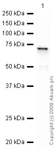
Anti-Galectin 8/Gal-8 antibody (ab42879) at 1 µg/ml + Galectin 8/Gal-8 Recombinant Protein at 0.1 µg
Secondary
Goat Polyclonal to rabbit IgG - H&L - Pre adsorbed (HRP) at 1/3000 dilution
Performed under reducing conditions.
Predicted band size: 35 kDa
Observed band size: 70 kDa why is the actual band size different from the predicted?ab42879 recognizes the full length tagged recombinant Galectin 8/Gal-8 protein which has an expected molecular weight of 65.5 kDa.
-
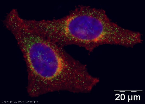
ICC/IF image of ab42879 stained human HeLa cells. The cells were PFA fixed (10 min), permabilised in 0.1% PBS-Tween (20 min) and incubated with the antibody (ab42879, 5µg/ml) for 1h at room temperature. 1%BSA / 10% normal goat serum / 0.3M glycine was used to block non-specific protein-protein interactions. The secondary antibody (green) was Alexa Fluor® 488 goat anti-rabbit IgG (H+L) used at a 1/1000 dilution for 1h. Alexa Fluor® 594 WGA was used to label plasma membranes (red). DAPI was used to stain the cell nuclei (blue).
Protocols
Datasheets and documents
References (0)
ab42879 has not yet been referenced specifically in any publications.
Images
-
Anti-Galectin 8/Gal-8 antibody (ab42879) at 1 µg/ml + Galectin 8/Gal-8 Recombinant Protein at 0.1 µg
Secondary
Goat Polyclonal to rabbit IgG - H&L - Pre adsorbed (HRP) at 1/3000 dilution
Performed under reducing conditions.
Predicted band size: 35 kDa
Observed band size: 70 kDa why is the actual band size different from the predicted?ab42879 recognizes the full length tagged recombinant Galectin 8/Gal-8 protein which has an expected molecular weight of 65.5 kDa.
-
ICC/IF image of ab42879 stained human HeLa cells. The cells were PFA fixed (10 min), permabilised in 0.1% PBS-Tween (20 min) and incubated with the antibody (ab42879, 5µg/ml) for 1h at room temperature. 1%BSA / 10% normal goat serum / 0.3M glycine was used to block non-specific protein-protein interactions. The secondary antibody (green) was Alexa Fluor® 488 goat anti-rabbit IgG (H+L) used at a 1/1000 dilution for 1h. Alexa Fluor® 594 WGA was used to label plasma membranes (red). DAPI was used to stain the cell nuclei (blue).


