Anti-Endostatin/COL18A1 antibody (ab3453)
")
Key features and details
- Rabbit polyclonal to Endostatin/COL18A1
- Suitable for: ICC/IF, IHC-P
- Reacts with: Mouse, Human, Non human primates
- Isotype: IgG
Overview
-
Product name
Anti-Endostatin/COL18A1 antibody
See all Endostatin/COL18A1 primary antibodies -
Description
Rabbit polyclonal to Endostatin/COL18A1 -
Host species
Rabbit -
Specificity
Detects recombinant human Endostatin/COL18A1.
-
Tested applications
Suitable for: ICC/IF, IHC-Pmore details -
Species reactivity
Reacts with: Mouse, Human, Non human primates -
Immunogen
Synthetic peptide corresponding to Human Endostatin/COL18A1 aa 129-142.
Sequence:RRLM/TESYCETWRTE
(Peptide available asab4983) -
General notes
ab3453 has been recombinant only tested in Western blot
This product was previously labelled as Endostatin


Properties
-
Form
Liquid -
Storage instructions
Shipped at 4°C. Store at +4°C short term (1-2 weeks). Upon delivery aliquot. Store at -20°C or -80°C. Avoid freeze / thaw cycle. -
Storage buffer
Preservative: 0.05% Sodium azide
Constituents: 0.1% BSA, 99% PBS -
 Concentration information loading...
 -
Purity
Immunogen affinity purified -
Clonality
Polyclonal -
Isotype
IgG -
Research areas
Images
-
Immunocytochemistry/ Immunofluorescence - Anti-Endostatin/COL18A1 antibody (ab3453)
ab3453 labelling Endostatin/COL18A1 (green) in 293T cells (right) compared with a negative control (left) by Immunocytochemistry/Immunofluorscence. Formalin-fixed cells were permeabilized with 0.1% Triton X-100 in TBS for 5-10 minutes, blocked with 3% BSA-PBS for 30 minutes at room temperature. Cells were incubated with the primary antibody (1:200 in 3% BSA-PBS) overnight at 4 ºC. A DyLight 488-conjugated Goat anti-rabbit IgG was used as the secondary antibody. Red (phalloidin) - F-actin, Blue (DAPI) - nuclei (blue). Images were taken at a magnification of 60x.
-
 Immunocytochemistry/ Immunofluorescence - Anti-Endostatin/COL18A1 antibody (ab3453)
ab3453 labelling Endostatin/COL18A1 (green) in A431 cells (right) compared with a negative control (left) by Immunocytochemistry/Immunofluorscence. Formalin-fixed cells were permeabilized with 0.1% Triton X-100 in TBS for 5-10 minutes, blocked with 3% BSA-PBS for 30 minutes at room temperature. Cells were incubated with the primary antibody (1:200 in 3% BSA-PBS) overnight at 4 ºC. A DyLight 488-conjugated Goat anti-rabbit IgG was used as the secondary antibody. Red (phalloidin) - F-actin, Blue (DAPI) - nuclei (blue). Images were taken at a magnification of 60x.
-
 Immunocytochemistry/ Immunofluorescence - Anti-Endostatin/COL18A1 antibody (ab3453)
ab3453 labelling Endostatin/COL18A1 (green) in NIH-3T3 cells (right) compared with a negative control (left) by Immunocytochemistry/Immunofluorscence. Formalin-fixed cells were permeabilized with 0.1% Triton X-100 in TBS for 5-10 minutes, blocked with 3% BSA-PBS for 30 minutes at room temperature. Cells were incubated with the primary antibody (1:200 in 3% BSA-PBS) overnight at 4 ºC. A DyLight 488-conjugated Goat anti-rabbit IgG was used as the secondary antibody. Red (phalloidin) - F-actin, Blue (DAPI) - nuclei (blue). Images were taken at a magnification of 60x.
-
 Immunohistochemistry (Formalin/PFA-fixed paraffin-embedded sections) - Anti-Endostatin/COL18A1 antibody (ab3453)
ab3453 labelling Endostatin/COL18A1 in the secretion of Human kidney tissue (right) compared with a negative control (left) by Immunohistochemistry (formalin/PFA-fixed paraffin embedded sections). To expose target proteins, antigen retrieval method was performed using 10mM sodium citrate (pH 6.0), microwaved for 8-15 min. Following antigen retrieval, tissues were blocked in 3% H2O2-methanol for 15 min at room temperature. Tissue sections were incuabted with the primary antibody (1:100 in 3% BSA-PBS) overnight at 4°C. A HRP-conjugated anti-rabbit IgG was used as the secondary antibody, followed by colorimetric detection using a DAB kit. Tissues were counterstained with hematoxylin and dehydrated with ethanol and xylene to prep for mounting.
-
 Immunohistochemistry (Formalin/PFA-fixed paraffin-embedded sections) - Anti-Endostatin/COL18A1 antibody (ab3453)
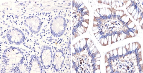
ab3453 labelling Endostatin/COL18A1 in the secretion of Human colon tissue (right) compared with a negative control (left) by Immunohistochemistry (formalin/PFA-fixed paraffin embedded sections). To expose target proteins, antigen retrieval method was performed using 10mM sodium citrate (pH 6.0), microwaved for 8-15 min. Following antigen retrieval, tissues were blocked in 3% H2O2-methanol for 15 min at room temperature. Tissue sections were incuabted with the primary antibody (1:200 in 3% BSA-PBS) overnight at 4°C. A HRP-conjugated anti-rabbit IgG was used as the secondary antibody, followed by colorimetric detection using a DAB kit. Tissues were counterstained with hematoxylin and dehydrated with ethanol and xylene to prep for mounting.
-
 Immunohistochemistry (Formalin/PFA-fixed paraffin-embedded sections) - Anti-Endostatin/COL18A1 antibody (ab3453)
ab3453 labelling Endostatin/COL18A1 in the secretion of Mouse kidney tissue (right) compared with a negative control (left) by Immunohistochemistry (formalin/PFA-fixed paraffin embedded sections). To expose target proteins, antigen retrieval method was performed using 10mM sodium citrate (pH 6.0), microwaved for 8-15 min. Following antigen retrieval, tissues were blocked in 3% H2O2-methanol for 15 min at room temperature. Tissue sections were incuabted with the primary antibody (1:100 in 3% BSA-PBS) overnight at 4°C. A HRP-conjugated anti-rabbit IgG was used as the secondary antibody, followed by colorimetric detection using a DAB kit. Tissues were counterstained with hematoxylin and dehydrated with ethanol and xylene to prep for mounting.


