Anti-ELMO1 antibody (ab2239)
Key features and details
- Goat polyclonal to ELMO1
- Suitable for: IHC-P, WB
- Reacts with: Mouse, Rat, Human
- Isotype: IgG
Overview
-
Product name
Anti-ELMO1 antibody
See all ELMO1 primary antibodies -
Description
Goat polyclonal to ELMO1 -
Host species
Goat -
Specificity
This antibody is expected to recognise both human isoforms. -
Tested applications
Suitable for: IHC-P, WBmore details -
Species reactivity
Reacts with: Mouse, Rat, Human
Predicted to work with: Dog
-
Immunogen
Synthetic peptide:
PKEPSNYDFVYDCN
, corresponding to C terminal amino acids 714-727 of Human ELMO1. -
General notes
The Life Science industry has been in the grips of a reproducibility crisis for a number of years. Abcam is leading the way in addressing this with our range of recombinant monoclonal antibodies and knockout edited cell lines for gold-standard validation. Please check that this product meets your needs before purchasing.
If you have any questions, special requirements or concerns, please send us an inquiry and/or contact our Support team ahead of purchase. Recommended alternatives for this product can be found below, along with publications, customer reviews and Q&As
Properties
-
Form
Liquid -
Storage instructions
Shipped at 4°C. Upon delivery aliquot and store at -20°C or -80°C. Avoid repeated freeze / thaw cycles. -
Storage buffer
pH: 7.30
Preservative: 0.02% Sodium azide
Constituents: Tris buffered saline, 0.5% BSA -
 Concentration information loading...
 -
Purity
Immunogen affinity purified -
Purification notes
Purified from goat serum by ammonium sulphate precipitation followed by antigen affinity chromatography using the immunizing peptide. -
Clonality
Polyclonal -
Isotype
IgG -
Research areas
Images
-
All lanes : Anti-ELMO1 antibody (ab2239) at 0.3 µg/ml
Lane 1 : Human Frontal Cortex cell lysate (35µg protein in RIPA buffer).
Lane 2 : Mouse cell lysate (35µg protein in RIPA buffer).
Lane 3 : Rat cell lysate (35µg protein in RIPA buffer).Primary incubation was 1 hour. Detected by chemiluminescence.
-
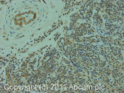
IHC image of ab2239 staining in human normal lymphoid formalin fixed paraffin embedded tissue section, performed on a Leica BondTM system using the standard protocol B. The section was pre-treated using heat mediated antigen retrieval with sodium citrate buffer (pH6, epitope retrieval solution 1) for 20 mins. The section was then incubated with ab2239, 10µg/ml, for 15 mins at room temperature and detected using an HRP conjugated compact polymer system. DAB was used as the chromogen. The section was then counterstained with haematoxylin and mounted with DPX.
For other IHC staining systems (automated and non-automated) customers should optimize variable parameters such as antigen retrieval conditions, primary antibody concentration and antibody incubation times.
-
Ab2239 staining (1
µ g/ml) of Jurkat lysate (RIPA buffer, 35µ g total protein per lane). Primary incubated for 1 hour. Detected by western blot using chemiluminescence. Ab2239 staining (1µg/ml) of Jurkat lysate (RIPA buffer, 35µg total protein per lane). Primary incubated for 1 hour. Detected by western blot using chemiluminescence.


