Anti-APC15 antibody (ab122349)
")
Key features and details
- Rabbit polyclonal to APC15
- Suitable for: WB, ICC/IF, IHC-P
- Reacts with: Human
- Isotype: IgG
Overview
-
Product name
Anti-APC15 antibody -
Description
Rabbit polyclonal to APC15 -
Host species
Rabbit -
Tested applications
Suitable for: WB, ICC/IF, IHC-Pmore details -
Species reactivity
Reacts with: Human -
Immunogen
Recombinant fragment corresponding to Human APC15 aa 6-79.
Sequence:PSLFPRVTETLWFNLDRPCVEETELQQQEQQHQAWLQSIAEKDNNLVPIG KPASEHYDDEEEED
-
Positive control
- Human appendix tissue.
-
General notes
The Life Science industry has been in the grips of a reproducibility crisis for a number of years. Abcam is leading the way in addressing this with our range of recombinant monoclonal antibodies and knockout edited cell lines for gold-standard validation. Please check that this product meets your needs before purchasing.
If you have any questions, special requirements or concerns, please send us an inquiry and/or contact our Support team ahead of purchase. Recommended alternatives for this product can be found below, along with publications, customer reviews and Q&As
Properties
-
Form
Liquid -
Storage instructions
Shipped at 4°C. Upon delivery aliquot and store at -20°C. Avoid freeze / thaw cycles. -
Storage buffer
pH: 7.20
Preservative: 0.02% Sodium azide
Constituents: 59% PBS, 40% Glycerol (glycerin, glycerine) -
 Concentration information loading...
 -
Purity
Immunogen affinity purified -
Clonality
Polyclonal -
Isotype
IgG -
Research areas
Images
-
Immunocytochemistry/ Immunofluorescence - Anti-APC15 antibody (ab122349)Immunofluorescent staining of Human cell line U-2 OS shows positivity in nucleus but not nucleoli and vesicles. Recommended concentration of ab122349 1-4 µg/ml. Cells treated with PFA/Triton X-100.
-
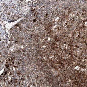 Immunohistochemistry (Formalin/PFA-fixed paraffin-embedded sections) - Anti-APC15 antibody (ab122349)
ab122349, at a 1/450 dilution, staining APC15 in paraffin-embedded Human appendix tissue by immunohistochemistry.
-
 Western blot - Anti-APC15 antibody (ab122349)Lane 1: Marker [kDa] 250; 130; 95; 72; 55; 36; 28; 17; 10
Lane 2: Negative control (vector only transfected HEK293T lysate)
Lane 3: Over-expression Lysate (Co-expressed with a C-terminal myc-DDK tag (~3.1 kDa) in mammalian HEK293T cells. Lane 1: Marker [kDa] 250; 130; 95; 72; 55; 36; 28; 17; 10
Lane 2: Negative control (vector only transfected HEK293T lysate)
Lane 3: Over-expression Lysate (Co-expressed with a C-terminal myc-DDK tag (~3.1 kDa) in mammalian HEK293T cells.


