ADP/ATP Ratio Assay Kit (Bioluminescent) (ab65313)
Key features and details
- Assay type: Semi-quantitative
- Detection method: Luminescent
- Platform: Microplate reader
- Assay time: 30 min
- Sample type: Adherent cells, Suspension cells, Tissue
- Sensitivity: 100 cells/well
Overview
-
Product name
ADP/ATP Ratio Assay Kit (Bioluminescent) -
Detection method
Luminescent -
Sample type
Tissue, Adherent cells, Suspension cells -
Assay type
Semi-quantitative -
Sensitivity
Assay time
0h 30mProduct overview
ADP/ATP Ratio Assay Kit (Bioluminescent) ab65313 is based on the bioluminescent detection of ADP and ATP levels. It can be used for a rapid screening of apoptosis, necrosis, growth arrest, and cell proliferation simultaneously in mammalian cells.
In the ADP/ATP assay protocol, luciferase catalyzes the conversion of ATP and luciferin to light, which in turn can be measured using a luminometer or Beta Counter. The ADP level is measured by its conversion to ATP that is subsequently detected using the same reaction.
The assay can be fully automatic for high throughput and is highly sensitive (detects 100 mammalian cells/well).
ADP/ATP Ratio assay protocol summary:
- transfer suspension cells, or nucleotide releasing buffer treated adherent cells, to plate
- add ATP reaction mix and incubate for 2 min
- analyze with luminescence plate reader to measure ATP
- after preparing ADP reaction mix and measuring luminescence levels again, add ADP reaction mix to same wells and incubate for 2 min
- analyze with luminescence plate reader to measure ADPNotes
Changes in the ADP/ATP ratio have been used to differentiate the different modes of cell death and viability. Increased levels of ATP and decreased levels of ADP have been recognized in proliferating cells. In contrast, decreased levels of ATP and increased levels of ADP are recognized in apoptotic cells. The decrease in ATP and increase in ADP are much more pronounced in necrosis than apoptosis.
Related assays
Review the cell health assay guide to learn about kits to perform a cell viability assay, cytotoxicity assay and cell proliferation assay.
Review the metabolism assay guide to learn about assays for metabolites, metabolic enzymes, mitochondrial function, and oxidative stress, and also about how to assay metabolic function in live cells using your plate reader.
Platform
Microplate readerProperties
-
Storage instructions
Store at -20°C. Please refer to protocols. -
Components Identifier 200 tests ADP Converting Enzyme (Lyophilised) Blue Cap 1 vial ATP Monitoring Enzyme (Lyophilised) 1 vial Enzyme Reconstitution Buffer 1 x 2.15ml Nucleotide Releasing Buffer 1 x 50ml -
Research areas
-
Relevance
The changes in ADP/ATP ratio have been used to differentiate the different modes of cell death and viability. Increased levels of ATP and decreased levels of ADP have been recognized in proliferating cells. In contrast, decreased levels of ATP and increased levels of ADP are recognized in apoptotic cells. The decrease in ATP and increase in ADP are much more pronounced in necrosis than apoptosis.
Properties
-
Storage instructions
Store at -20°C. Please refer to protocols. -
Components Identifier 200 tests ADP Converting Enzyme (Lyophilised) Blue Cap 1 vial ATP Monitoring Enzyme (Lyophilised) 1 vial Enzyme Reconstitution Buffer 1 x 2.15ml Nucleotide Releasing Buffer 1 x 50ml -
Research areas
-
Relevance
The changes in ADP/ATP ratio have been used to differentiate the different modes of cell death and viability. Increased levels of ATP and decreased levels of ADP have been recognized in proliferating cells. In contrast, decreased levels of ATP and increased levels of ADP are recognized in apoptotic cells. The decrease in ATP and increase in ADP are much more pronounced in necrosis than apoptosis.
Images
-
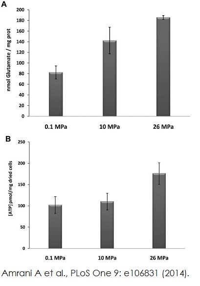
 Functional studies - ab65313 Image from Amrani A et al., PLoS One 9(9). Fig 5A. doi: 10.1371/journal.pone.0106831 Reproduced under the Creative Commons license http://creativecommons.org/licenses/by/4.0/
Quantitation of glutamate levels (A) using ab138883 and intracellular ATP (B) using ab65313 in D. hydrothermalis cells grown under different pressure conditions.
-
 Functional studies - ab65313 Bhattacharyya S.,PLoS One 8(11), Fig 5d & f. doi: 10.1371/journal.pone.0079167 Reproduced under the Creative Commons license http://creativecommons.org/licenses/by/4.0/
ADP/ATP ratio in cystathionine-beta-synthase slienced A2780 (Ovarian cancer cell line) cells and AOAA (aminooxyacetic acid) treated A2780 cells were measured using ADP/ATP ratio assay kit (ab65313).
-
 Functional studies - ab65313 Image from Julien S.G., PLoS Biol 15(2), Fig 8C. doi: 10.1371/journal.pbio.1002597. Reproduced under the Creative Commons license http://creativecommons.org/licenses/by/4.0/
ADP/ATP ratio were examined in both narciclasine (ncls) and vehicle (veh) treated C2C12 myotubes with or without PA treatment using ADP/ATP ratio assay kit (ab65313).


